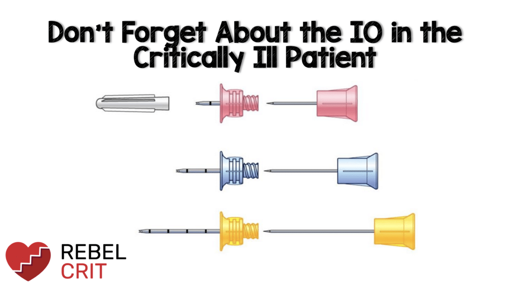
In these situations, the American Heart Association (AHA) and the European Resuscitation Council (ERC) of 2015 recommend the intraosseous (IO) route after the peripheral route and before the central venous route (1). However, this is often overlooked in emergencies whether from lack of access to IO supplies, lack of training by health care staff, or not being considered as an alternative means of access during the medical decision-making process.
Kristin Wiley DO 1, Frank Lodeserto MD2
Affiliations
- Department of Emergency Medicine, Cape Fear Valley Medical Center, Fayetteville, North Carolina
- Department of Critical Care Medicine, Cape Fear Valley Medical Center, Fayetteville, North Carolina
IO vs. CVC vs PIV
The critically ill patient is often associated with pathology that makes IV access difficult such as shock state, hypovolemia, obesity, IV drug abuse, end-stage renal disease, cardiac arrest, as well as other conditions. Studies have compared IO to peripheral intravenous (PIV) and central venous (CVC) access for resuscitation. One meta-analysis in trauma patients demonstrated that the first-attempt success rate was higher in IO compared to PIVs (2).
Many clinicians don’t consider IO placement while others consider it a last resort or only a pre-hospital procedure. The IO can actually be your “GO-TO” emergency line or your second choice after failed PIV access. Several studies have shown IO placement times <20 seconds (3) but usually less than 1 minute (3-6), near 100% first-pass success rate (3-7), similar flow rates to that of a central line (6), and little or no complications. Once you have secured IO access at one or even multiple sites, and begun your resuscitation, then you may have more time to secure sterile central access if still indicated.
The “crash” or “dirty” central line, which forgoes strict sterile precautions, should be the last consideration in resuscitation as it increases risks for bloodstream infections, has more associated complications, and takes a potential access site required for ECMO cannulation, transvenous pacemaker, or other cardiac devices. If central line placement is indicated it should be performed once the patient is adequately resuscitated under sterile precautions.
Studies reviewed landmark-based CVC compared to IO; using IJ, subclavian, and femoral CVC sites. CVC placement took significantly longer to place, had a higher first-pass fail rate, and increased complications compared to IO placement (7-8). A study by Lee et al (7) compared femoral CVC placement to IO and demonstrated a first-pass success pass rate with IO of 90.3% compared to only 37.5% in the CVC group. This study also showed the median time for IO placement was only 1.2 minutes compared to a mean placement time of 10.7 minutes CVC group.
The current standard of practice has moved away from landmark-based central line placement given the efficacy and safety of ultrasound-based techniques. We were not able to identify a study specifically comparing US-guided CVC compared to IO placement. One may speculate that the US-guided CVC placement would have a higher first-pass success rate with fewer complications, however, this may potentially add time to the procedure depending on the operator and institution’s use of ultrasound during emergencies and maintaining sterile technique with the US probe.
In cardiac arrest, a delay in IV access subsequently results in a delay in epinephrine administration. Given AHA guidelines recommend early epinephrine administration in cardiac arrest, obtaining access is imperative for resuscitative efforts (9) When evaluating over 300,000 out-of-hospital cardiac arrests, a meta-analysis found no significant change in primary outcomes when IV and IO access were compared (10) . For in-hospital cardiac arrest, IO was shown to be non-inferior to PIV access in overall survival and neurologic status and should be used as an alternative to PIV until more definitive access routes can be performed (11) IO cannulation has shown to be a rapid, safe way to easily administer drugs, fluids, and
blood products in the acute setting (12).
IO Location, Contraindications & Infusion Rates
IO access can be obtained in numerous locations with the most common sites being the proximal humerus and proximal tibia. The standard IO is a 15-gauge, short, non-collapsible access while a typical triple-lumen CVC has two 18-gauge and one 16-gauge lumen, both with long catheters, resulting in slower flow rates. The humeral IO can achieve flow rates of up to 5L/hour and given closer proximity to central circulation can reach the right atrium in ~ 3 seconds. The tibial IO can achieve fluid rates of 1L/h with pressure bag (13). The proximal tibial site has less risk of dislodgement during resuscitation and is easier to obtain in obese patients. Additional sites such as the sternal, iliac crest, distal femur, and distal tibia have also been utilized, and the use of these alternative sites should be considered based on the clinical presentation.
Contraindications to IO placement include:
• Skin infection or cellulitis at the insertion site
• Fractured bone proximal to IO insertion
• Severe underlying bone disease (osteogenesis imperfecta, osteoporosis, osteomyelitis, compartment syndrome)
• Burns
• A recent failed IO attempt in the same bone
IO access can be complicated by extravasation, bone injury, soft tissue necrosis, osteomyelitis,
and osteo-fascial compartment syndrome. Extravasation is notably the most common
complication and the overall complication rates are very rare.
Bottom Line
• All emergency medications can be given through an IO
• IO can be used in all emergencies including trauma and cardiac arrest.
• IO is faster and safer to place than a CVC and often faster than a PIV.
• IO requires minimal training and experience for placement.
• Flow rates in IO can be faster or at least comparable to that of a CVC.
• Labs can be drawn from an IO to help with rapid diagnosis in emergencies.
References:
1 Astasio-Picado Á et al. Clinical Management of Intraosseous Access in Adults in Critical Situations for Health Professionals. Healthcare. 2022 PMID: 35206981
2. Wang, D. et al. Efficacy of intraosseous access for trauma resuscitation: a systematic review and meta-analysis. World J Emerg Surg 2023 PMID: 36918947
3. Ngo AS, Oh JJ, Chen Y, Yong D, Ong MEH. Intraosseous vascular access in adults using the EZ-IO in an emergency department. Int J Emerg Med 2009 PMID: 20157465
4. Ong MEH, Chan YH, Oh JJ, et al. An observational prospective study comparing tibial and humeral intraosseous access using the EZ-IO. Am J Emerg Med PMID: 19041528
5. Iserson KV et al. Intraosseous infusions in adults. J Emerg Med 1989. PMID: 2625519
6. Iwama H. et al. Emergency fields, obtaining intravascular access for cardiopulmonary arrest patients is occasionally difficult and time-consuming. J. Trauma 1996. PMID: 8913236
7 Lee PM et al. Intraosseous versus central venous catheter utilization and performance during inpatient medical emergencies. Crit Care Med. 2015 PMID: 25768683
8 Leidel BA. Et al. Comparison of intraosseous versus central venous vascular access in adults under resuscitation in the emergency department with inaccessible peripheral veins. Resuscitation. 2012 PMID: 21893125.
9 Zhang W et al. Intravenous vs intraosseous adrenaline administration in cardiac arrest: A protocol for systematic review and meta-analysis. Medicine (Baltimore). 2020. PMID: 33350794
10 Meilandt C. et al. Intravenous vs.intraosseous vascular access during out-of-hospital cardiac arrest – protocol for a randomized clinical trial. Resusc Plus. 2023 PMID: 37502742
11 Schwalbach KT et al. Impact of intraosseous versus intravenous resuscitation during in-hospital cardiac arrest: A retrospective study. Resuscitation. 2021 PMID: 34273470
12 Petitpas F et al. Use of intra-osseous access in adults: a systematic review. Crit Care. 2016 PMID:27075364
13 Astasio-Picado Á et al. Clinical Management of Intraosseous Access in Adults in Critical Situations for Health Professionals. Healthcare 2022 PMID: 35206981
Post Peer Reviewed By: Frank Lodeserto, MD & Salim R. Rezaie, MD (Twitter/X: @srrezaie)


