Remove ecg cardiac-memory
article thumbnail

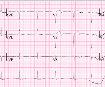
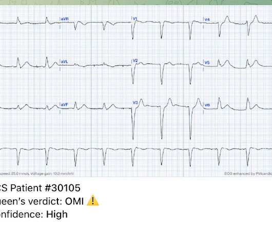
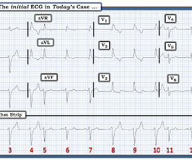
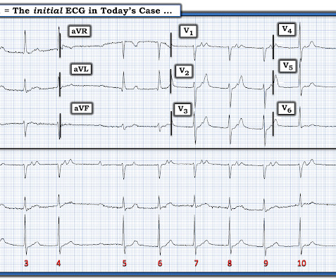
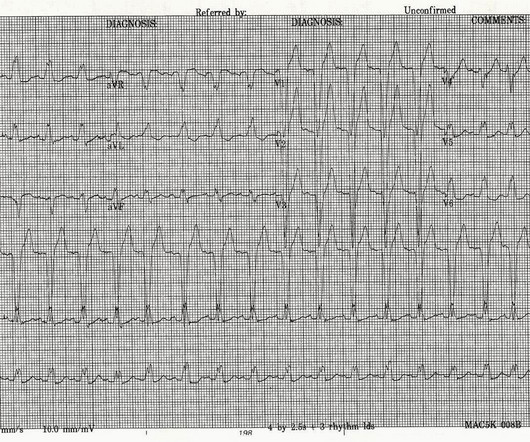
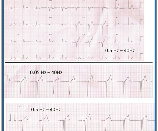
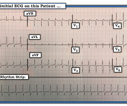
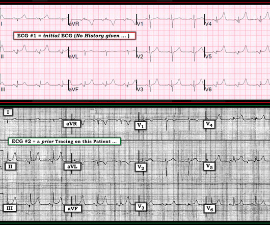
Explain this ECG in the context of active chest pain, slightly elevated troponin without a delta, RCA culprit, and previous with LBBB

Dr. Smith's ECG Blog

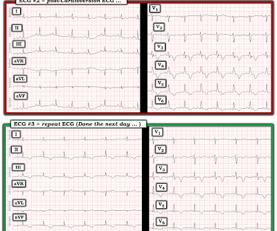
It turns out that she spends much of her time in LBBB (see ECGs below) What is going on??? Cardiac Memory: After conversion from LBBB or Paced Rhythm back to normal conduction, T-wave inversion is common and is called "Cardiac Memory" See this post and explanation: Chest pain and LBBB. link] Shvilkin et al.

EKG/ECG 74
article thumbnail

ECG Blog #386 — OMI or Something Else?

Ken Grauer, MD

The ECG in Figure-1 was obtained from a previously healthy middle-aged man — who while performing his regular exercise routine, developed "slight" chest discomfort and "palpitations". Surprisingly — he was hemodynamically stable with minimal discomfort at the time his initial ECG in Figure-1 was recorded. Could the patient have WPW?

EKG/ECG 195
Insiders

Sign Up for our Newsletter

This site is protected by reCAPTCHA and the Google Privacy Policy and Terms of Service apply.

Trending Sources

article thumbnail

An 80 year old woman with Left Bundle Branch Block (LBBB) and pleuritic chest pain

Dr. Smith's ECG Blog

This was the ECG obtained at triage. This ECG was recorded and was reviewed remotely by a cardiologist: What do you think? The Queen of Hearts agrees: Here the Queen explains why: However, it was not interpreted correctly by the providers: ED interpretation of ECG: "paced rhythm, LBBB but no STEMI pattern." Next trop in AM.

EKG/ECG 79
article thumbnail

ECG Blog #373 — 86yo and this Rhythm.

Ken Grauer, MD

The 12-lead ECG and long lead II rhythm strip in Figure-1 was obtained from an 86-year old man — who presented to the ED ( E mergency D epartment ) with presyncope. How would YOU interpret the ECG in Figure-1 ? Figure-1: The initial ECG in today's case — obtained from an 86-year old man with presyncope, but no chest pain. (

EKG/ECG 195
article thumbnail

ECG Blog #362 — Funny Rhythm in a 14-Year Old.

Ken Grauer, MD

Cardiac monitoring revealed a series of interesting ( albeit asymptomatic ) rhythms, 2 of which are shown below in Figure-1. MY Approach to the Rhythm in Figure-1: As always — I favor a systematic approach to rhythm interpretation — with use of the P s, Q s, 3 R Memory Aid ( See ECG Blog #185 for more on the Ps, Qs, 3Rs ).

EKG/ECG 195
article thumbnail

Is there Terminal QRS Distortion?

Dr. Smith's ECG Blog

This ECG has ST Elevation in multiple leads. This ECG is interesting because it mimics TQRSD. This is a normal ECG. This is a great demonstration that a normal ECG, if there is no S-wave in V2 and/or V3, the lead without an S-wave MUST have a J-wave in order to be truly normal. Is it normal STE? Or is it ischemic STE?

EKG/ECG 52
article thumbnail

A Covid patient with cough and Fever. Why does the ST-T wave look so abnormal?

Dr. Smith's ECG Blog

I was reading through a stack of ECGs and saw this one. It turns out that this patient has cardiac amyloidosis. The ECG in today’s case is one in which Dr. Smith immediately knew he needed to find out what was going on with this patient! It turned out the patient had cardiac amyloidosis. What is going on here?

EKG/ECG 52