Remove ecg left-anterior-hemiblock
article thumbnail

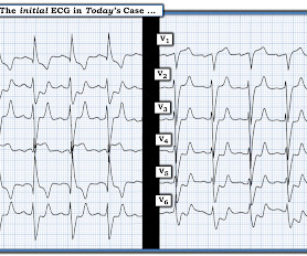
ECG Blog #430 — Just a Regular LBBB ECG?

Ken Grauer, MD

The ECG in Figure-1 — was obtained from an older man who had just completed dialysis — and , is now complaining of abdominal discomfort that radiates to his chest. Figure-1: The initial ECG in today's case. ( To improve visualization — I've digitized the original ECG using PMcardio ). How would YOU approach this case?

EKG/ECG 374
article thumbnail

ECG Blog #417 — AFib with Aberrancy?

Ken Grauer, MD

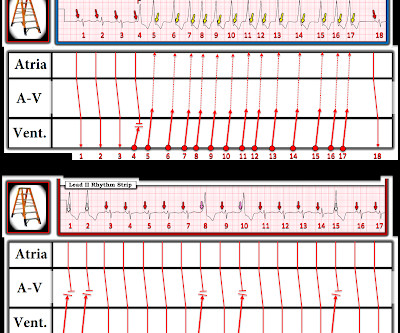
The ECG in Figure-1 was obtained from a previously healthy middle-aged man — who presented to the ED ( E mergency D epartment ) for shortness of breath. QUESTIONS: How would YOU interpret the ECG shown in Figure-1 ? Figure-1: The initial ECG in today's case. Figure-1: The initial ECG in today's case.

EKG/ECG 312
Insiders

Sign Up for our Newsletter

This site is protected by reCAPTCHA and the Google Privacy Policy and Terms of Service apply.

Trending Sources

article thumbnail

Explain this ECG in the context of active chest pain, slightly elevated troponin without a delta, RCA culprit, and previous with LBBB

Dr. Smith's ECG Blog

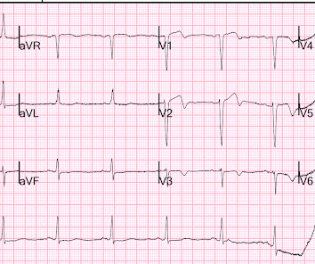
Pain is described as dull and pressure-like over the retrosternal area radiating to her neck with associated tingling of the left hand. Time 17 minutes Not much different One month earlier This is Left Bundle Branch Block (LBBB) without any sign of ischemia. It is rated 8/10 in severity. She has associated shortness of breath.

EKG/ECG 74
article thumbnail

A fascinating electrophysiology case. What is this wide complex tachycardia, and how best to manage it?

Dr. Smith's ECG Blog

The ECG below was recorded. The ECG was interpreted as showing atrial flutter with 2:1 conduction. Answer : The ECG above shows a regular wide complex tachycardia. Said differently, the ECG shows a rather slow ventricular tachycardia with a 2:1 VA conduction. How do you interpret the below ECG? There is some STE.

EKG/ECG 86
article thumbnail

60-something with wide complex tachycardia: from where does the rhythm originate?

Dr. Smith's ECG Blog

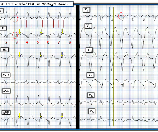
Here is her ED ECG: Here is the ED physician's interpretation: IMPRESSION UNCERTAIN REGULAR RHYTHM, wide complex tachycardia, likely p-waves. LEFT BUNDLE BRANCH BLOCK [120+ ms QRS DURATION, 80+ ms Q/S IN V1/V2, 85+ ms R IN I/aVL/V5/V6] Comparison Summary: LBBB and tachycardia are new. Figure-1: The 2 ECGs in this case ( See text ).

EKG/ECG 52
article thumbnail

A 30-something with palpitations and lightheadedness

Dr. Smith's ECG Blog

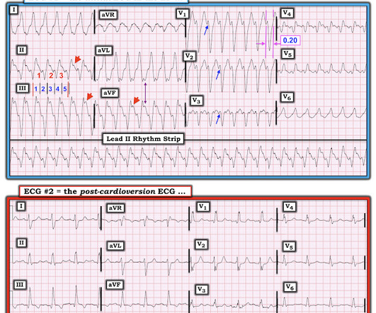
Here is his ED ECG: Regular Wide Complex Tachycardia at a rate of 229 First : What do you want to do? Next : What do you think is the ECG diagnosis? ECG analysis: T his is a regular wide complex tachycardia. Here is the post cardioversion ECG (texted to me): This what I texted back: "Could be WPW. Antidromic WPW).

EKG/ECG 52