article thumbnail

ECG Blog #415 — The Cath showed NO Occlusion!

Ken Grauer, MD

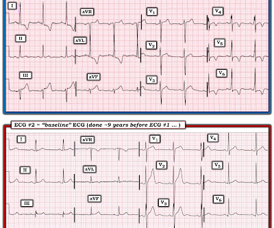
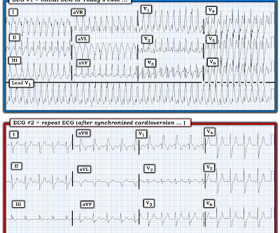
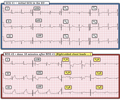
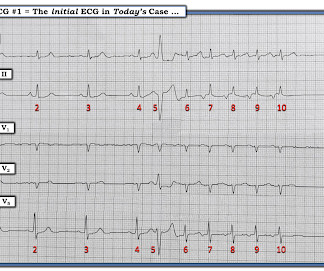
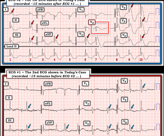
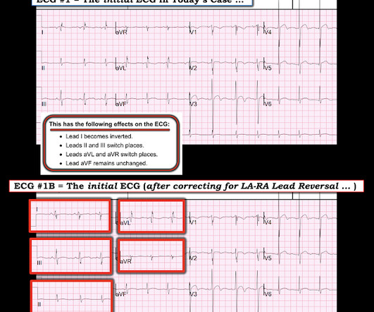
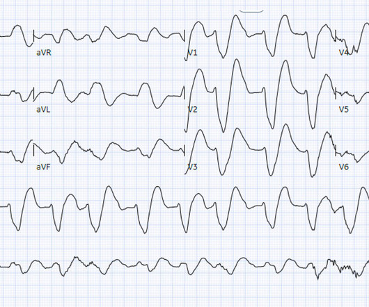
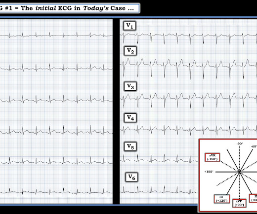
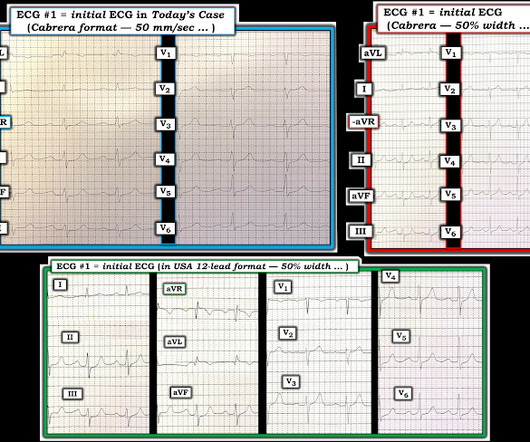
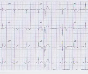
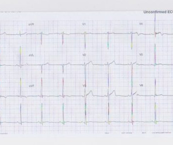
The ECG in Figure-1 was obtained following successful resuscitation. QUESTIONS: In view of the above history — How would YOU interpret the ECG in Figure-1 ? Is this ECG finding present in today’s initial ECG? Figure-1: The initial ECG in today's case — obtained after successful resuscitation from cardiac arrest. (

EKG/ECG 393
article thumbnail

ECG Blog #406 — To Do Additional Leads?

Ken Grauer, MD

For full discussion of this case — See ECG Blog #351 — == The ECG in Figure-1 — was obtained from a previously healthy older man who contacted EMS ( E mergency M edical S ervices ) because of "chest tightness" that began ~1 hour earlier. Given this history: QUESTIONS: How would YOU interpret the ECG in Figure-1 ?

EKG/ECG 375
Insiders

Sign Up for our Newsletter

This site is protected by reCAPTCHA and the Google Privacy Policy and Terms of Service apply.

Trending Sources

article thumbnail

ECG Blog #424 — Proportionality and the "Cut Off"

Ken Grauer, MD

The ECG in Figure-1 was obtained from a middle-aged woman — who presented with low back pain, shortness of breath and marked hypertension — but no CP ( C hest P ain ). QUESTIONS: In view of this history — How would YOU interpret this ECG? Figure-1: The initial ECG in today's case. Figure-1: The initial ECG in today's case.

EKG/ECG 231
article thumbnail

ECG Blog #412 — Is Cardiac Cath Indicated?

Ken Grauer, MD

The ECG in Figure-1 was obtained from a middle-aged man with known hypertension — who presented to the ED ( E mergency D epartment ) for CP ( C hest P ain ) over the preceding 2-3 days. QUESTIONS: How would YOU interpret the ECG in Figure-1 ? The Initial ECG in Today’s CASE: The rhythm in ECG #1 is sinus at 65-70/minute.

EKG/ECG 379
article thumbnail

ECG Video Blog #407 (292): Why the Patient Died?

Ken Grauer, MD

For full discussion of this case — See ECG Blog #292 — == The 2 ECGs shown in Figure-1 were obtained from a man in his 30s — who presented to the ED ( E mergency D epartment ) with chest pain that began several hours earlier. ECG #2 was recorded 1 hour after ECG #1. (22:3 0 min. Initial troponin was negative.

EKG/ECG 352
article thumbnail

Concerning EKG with a Non-obstructive angiogram. What happened?

Dr. Smith's ECG Blog

It appears EMS obtained two EKGs, but unfortunately these were not saved in the medical record. The EMS crew was only BLS certified, so EKG interpretation is not within their scope of practice. The patient arrived just after 10 AM, and the following EKG was obtained. There are no further EKGs or troponin measurements.

EKG/ECG 104
article thumbnail

ECG Blog #409 — Every-Other-Beat.

Ken Grauer, MD

The ECG in Figure-1 — was obtained from a patient with palpitations. Figure-1: The initial ECG in today's case. My Interpretation of the ECG in Figure-1: Since the patient is hemodynamically stable — there is time for systematic assessment of the rhythm. Figure-2: I've labeled the initial ECG in today's case. Figure-2 ).

EKG/ECG 430