Ken Grauer, MD

article thumbnail

ECG Blog #427 — To Cath this Elderly Patient?

Ken Grauer, MD

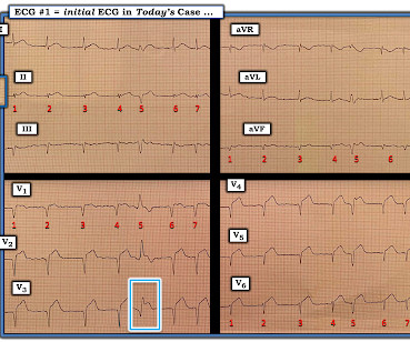
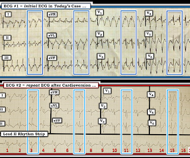
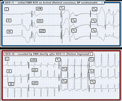
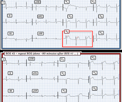
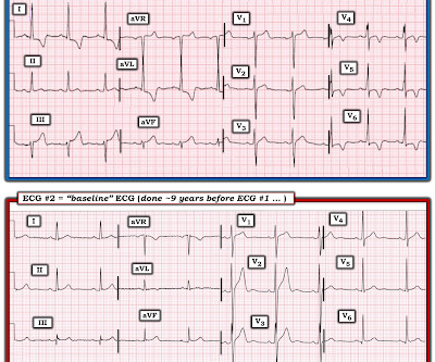
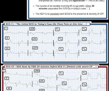
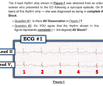
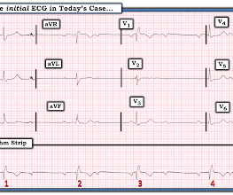
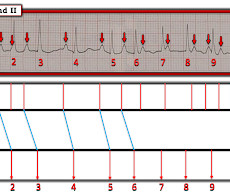
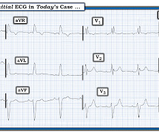
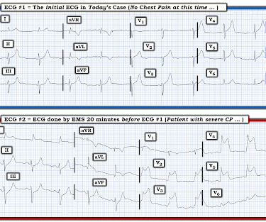
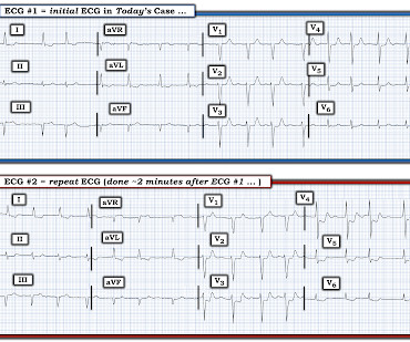
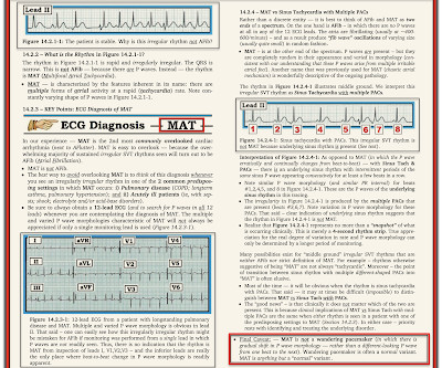
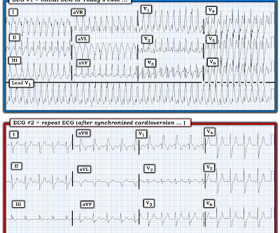
The ECG in Figure-1 — was obtained on the scene by EMS ( E mergency M edical S ervices ). The patient was a man in his 90s, who ~1 hour earlier, noted the onset of severe CP ( C hest P ain ). He was hemodynamically stable — but clearly distressed with a sense of “impending doom” at the time ECG #1 was recorded. Despite the patient’s age — he was independent, lived alone, had good mental function — and had family support.

EKG/ECG 298
article thumbnail

ECG Blog #426 — Are STEMI Criteria Met?

Ken Grauer, MD

The ECG in Figure-1 — was obtained from a 70-ish year old man with episodic CP ( C hest P ain ) over the previous 2-3 days , being awakened from sleep now for a more severe CP episode. QUESTIONS: In view of this history — How would YOU interpret this ECG? Should you activate the cath lab? Figure-1: The initial ECG in today's case. MY Initial Thoughts on Today's CASE: Although it is difficult from the brief history we are given, to determine the true onset of whatever might be happening — the pat

EKG/ECG 344
Insiders

Sign Up for our Newsletter

This site is protected by reCAPTCHA and the Google Privacy Policy and Terms of Service apply.

Trending Sources

article thumbnail

ECG Blog #425 — Are there P Waves?

Ken Grauer, MD

I was sent the ECG in Figure-1 — told only that the patient was 70 years old, and had a history of an ASD ( A trial S eptal D efect ). Serum K+ was normal. The patient was hemodynamically stable with ECG #1. QUESTIONS: How would YOU interpret the rhythm in Figure-1 ? Are there P waves? Figure-1: The initial ECG in today's case. MY Initial Thoughts on Today's CASE: Knowing that today's patient has a history of an ASD ( A trial S eptal D efect ) — is relevant to the interpretation of today's inter

EKG/ECG 281
article thumbnail

ECG Blog #424 — Proportionality and the "Cut Off"

Ken Grauer, MD

The ECG in Figure-1 was obtained from a middle-aged woman — who presented with low back pain, shortness of breath and marked hypertension — but no CP ( C hest P ain ). QUESTIONS: In view of this history — How would YOU interpret this ECG? Are the large, peaked T waves ( especially in lead V2 ) — likely to indicate hyperacute deWinter T waves? Figure-1: The initial ECG in today's case.

EKG/ECG 229
article thumbnail

ECG Blog #422 — Was Clubbing an ECG Hint?

Ken Grauer, MD

I was sent the ECG in Figure-1 — with the following history: The patient is a young man in his early 20s — who presents to the ED ( E mergency D epartment ) because of SOB ( S hortness O f B reath ) that had been ongoing for several hours. No chest pain. He reports a number of similar previous episodes over the past few years ( although apparently has not been formerly evaluated for this ).

EKG/ECG 402
article thumbnail

ECG Blog #421 — Has there been a Recent MI?

Ken Grauer, MD

What if you were asked to interpret the ECG in Figure-1 ? How would YOU interpret the rhythm? Even without the benefit of any history — Has there been a recent MI? Figure-1: The initial ECG in today's case. ( To improve visualization — I've digitized the original ECG using PMcardio ). MY Approach to Today’s Tracing: As always — I favor beginning assessment with a quick look at the long lead rhythm strips at the bottom of the tracing.

EKG/ECG 352
article thumbnail

ECG Blog #420 — A "Fast" Complete Heart Block?

Ken Grauer, MD

I was asked to interpret the 2-lead rhythm strip shown in Figure-1 — without the benefit of any history. What are YOUR thoughts? Is there AV block? If so — Is it complete AV block? Figure-1: You are asked to interpret this 2-lead rhythm strip without the benefit of any history. = NOTE: Today's rhythm is challenging — especially if you have not seen this type of rhythm before.

EKG/ECG 322