Remove blog
article thumbnail

ECG Blog #430 — Just a Regular LBBB ECG?

Ken Grauer, MD

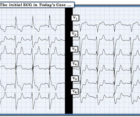
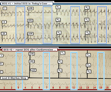
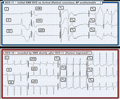
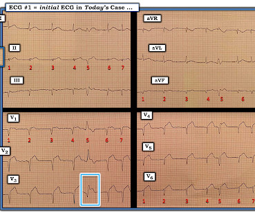
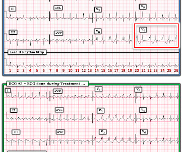
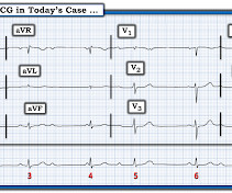
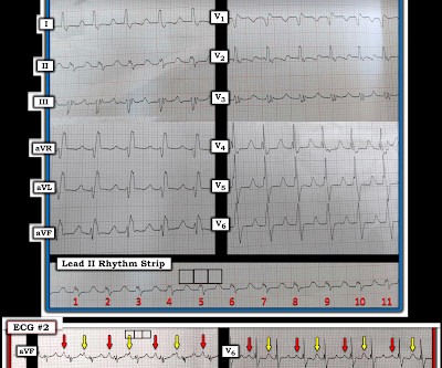
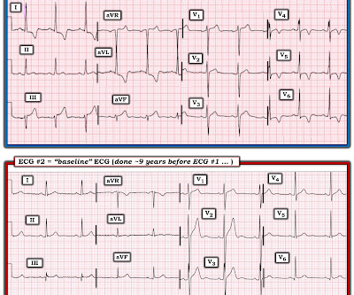
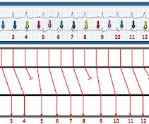
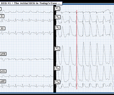
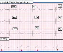
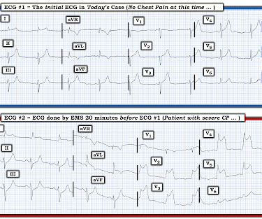
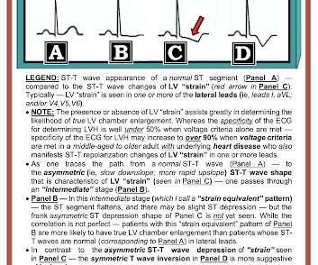
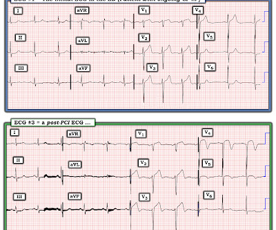
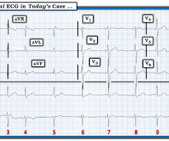
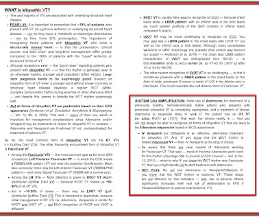
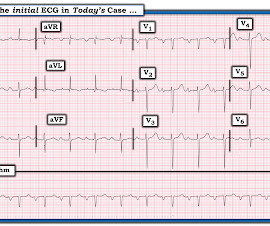
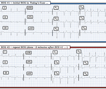
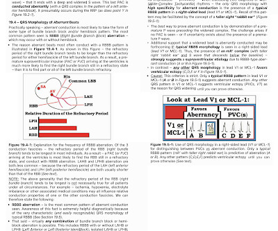
As I review in ECG Blog #204 — “typical” LBBB is characterized by a supraventricular rhythm with QRS widening, in which there is a monophasic R wave in left-sided leads I and V6 — and an all-negative ( or almost all negative ) QRS in right-sided lead V1. ECG Blog #294 — Reviews how to tell IF the " culprit " artery has reperfused.

EKG/ECG 374
article thumbnail

ECG Blog #422 — Was Clubbing an ECG Hint?

Ken Grauer, MD

This leaves us with the usual differential diagnosis for this rhythm presentation ( as per ECG Blog #361 ) ==> We need to consider i ) VT until proven otherwise: ii ) SVT with either preexisting BBB or aberrant conduction; — or , iii ) Something else ( ie, WPW, hyperkalemia, some other toxicity, etc. ). What Kind of Conduction Defect?

EKG/ECG 447
Insiders

Sign Up for our Newsletter

This site is protected by reCAPTCHA and the Google Privacy Policy and Terms of Service apply.

Trending Sources

article thumbnail

ECG Blog #426 — Are STEMI Criteria Met?

Ken Grauer, MD

As is also emphasized often in this ECG Blog — spontaneous reperfusion of the "culprit" artery is common — and, IF this occurs before a 2nd ECG is done, ST-T wave changes may "look better" ( See References to related Blog posts below ). ECG Blog #294 — Reviews how to tell IF the " culprit " artery has reperfused.

EKG/ECG 396
article thumbnail

ECG Blog #431 — My New ECG-Rhythm Podcasts!

Ken Grauer, MD

Easy LINKS — tinyurl.com/KG-ECG-Podcasts — [link] — Other ECG Audio PEARLS I previously made for my ECG Blog can be found in the right column of each page on this blog just below this icon — under, "ECG Audio PEARLS". I recently recorded a series of 4 podcasts regarding KEY concepts in ECG interpretation.

EKG/ECG 277
article thumbnail

ECG Blog #419 — The Cause of ECG #1?

Ken Grauer, MD

PEARL # 2: As described in ECG Blog #394 — QRS widening in the presence of sinus rhythm, in which QRS morphology is consistent with RBBB conduction in the chest leads — but LBBB conduction in the limb leads ( especially with a leftward axis ) — suggests the entity known as MBBB ( M asquerading B undle B ranch B lock ).

EKG/ECG 471
article thumbnail

ECG Blog #427 — To Cath this Elderly Patient?

Ken Grauer, MD

An example of a case in which the diagnosis of acute OMI was made purely by assessment of ST-T wave morphology in a PVC can be found HERE ( See My Comment at the bottom of this page in the October 8, 2018 post in Dr. Smith's ECG Blog ). = ECG Blog #294 — Reviews how to tell IF the " culprit " artery has reperfused.

EKG/ECG 348
article thumbnail

ECG Blog #409 — Every-Other-Beat.

Ken Grauer, MD

By the P s, Q s, 3 R Approach ( See ECG Blog #185 ): The rhythm is fast and QRS complexes are R egular. PEARL # 4: As emphasized in ECG Blog #204 , in which I review derivation of the bundle branch blocks — RBBB is a terminal conduction delay. ECG Blog #185 — Reviews the P s, Q s, 3 R Approach to Rhythm Interpretation.

EKG/ECG 430