Remove ecg preexcitation
article thumbnail

ECG Blog #371 — Palpitations Since Childhood.

Ken Grauer, MD

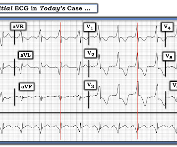
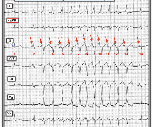
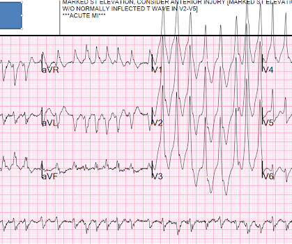
The ECG in Figure-1 is from a man in his 30s — who overall has been healthy, except for a history of "intermittent palpitations" that he has had since childhood. He was hemodynamically stable with ECG #1. Figure-1: The initial ECG in today's case. To improve visualization — I've digitized the original ECG using PMcardio ).

EKG/ECG 195
article thumbnail

Is all this "ST Depression" due to ischemia?

Dr. Smith's ECG Blog

Written by Magnus Nossen, with some edits by Smith This ECG was transmitted electronically by EMS for evaluation. How would you interpret the ST changes seen in this ECG? Will you accept this patient for emergent coronary angiogram based on the ECG changes? Does the ECG represent STEMI-negative OMI findings?

EKG/ECG 62
Insiders

Sign Up for our Newsletter

This site is protected by reCAPTCHA and the Google Privacy Policy and Terms of Service apply.

Trending Sources

article thumbnail

What do you think of this "Ventricular Bigeminy"?

Dr. Smith's ECG Blog

1] If we look at the patient’s previous EKG on record, we’re able to see that this pattern is not new: Interestingly, the patient does appear to have PVCs in this ECG as well as WPW beats. On the previous ECG, the diagnosis was missed, as it frequently is! I thought the ECG diagnosis was obvious. But many missed it. As per Drs.

EKG/ECG 52
article thumbnail

Chest pain and anterior ST depression. What’s the cause(s)?

Dr. Smith's ECG Blog

There were no prior ECGs, the first ECG was signed off as unremarkable, and the patient waited to be seen. An hour later the first troponin returned at 49 ng/L (normal <16 in females and <26 in males), so the patient was brought into an acute bed and the ECG was repeated, with ongoing symptoms. What do you think?

EKG/ECG 52
article thumbnail

Right Bundle Branch Block and Posterior OMI????

Dr. Smith's ECG Blog

mm) in V2 and V3, suggestive of RBBB with posterior OMI Whenever you see abnormal ST-T (ST elevation, ST depression, hyperacute T-waves), you MUST look at the entire ECG (rhythm, rate, P-waves, intervals, and QRS) to see if there is some abnormality among these which can explain the ST-T. Would you have recognized the acute posterior OMI?

EKG/ECG 52
article thumbnail

A woman in her 60s with palpitations

Dr. Smith's ECG Blog

The ECG there reportedly showed an irregular tachycardia, and the patient was immediately referred to the emergency room. Here is her ECG on arrival: There is a wide complex tachycardia that is irregularly irregular (this is difficult to determine at these very high rates). Figure-1: The initial ECG in today's case.

EKG/ECG 52
article thumbnail

Should we activate the cath lab? A Quiz on 5 Cases.

Dr. Smith's ECG Blog

Triage is backed up, and 10 minutes into your shift one of the ED nurses brings your several ECG s that has not been overread by a physician. ECG#1 ECG#2 ECG#3 ECG#4 ECG#5 See outcomes of all 5 below, with the Queen of Hearts AI Bot interpretation. (THE True Positive ECG#2 : Also sinus rhythm.