Remove ecg wide-qrs
article thumbnail

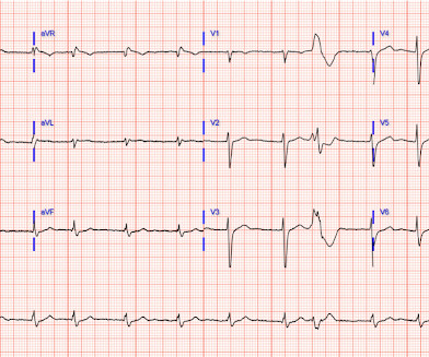
Instructors' Collection ECG: Regular Really Wide QRS Tachycardia

ECG Guru

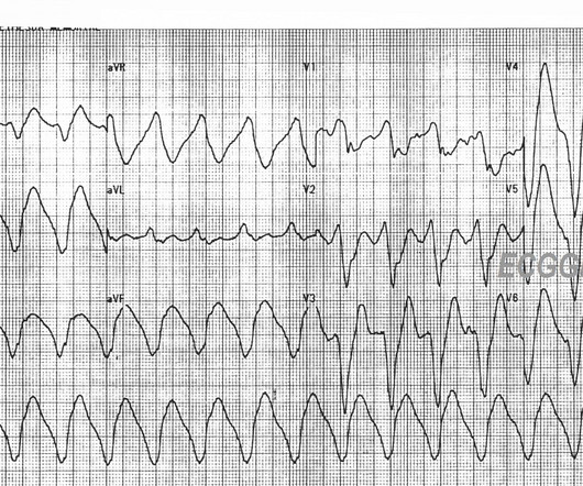
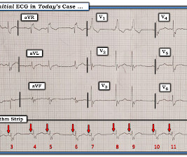
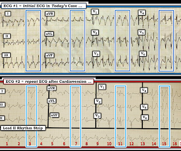
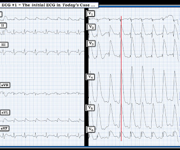
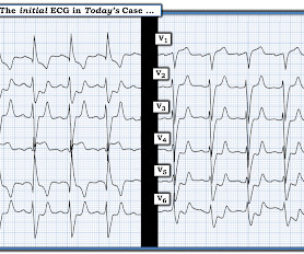
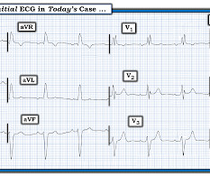
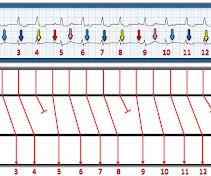
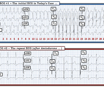
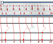
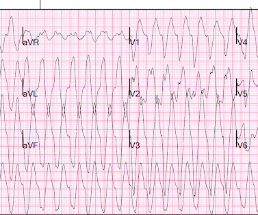
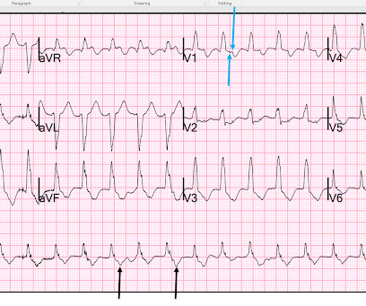
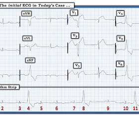
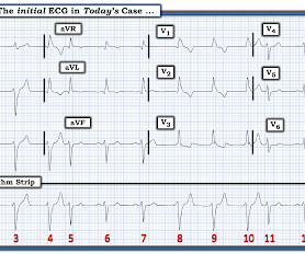
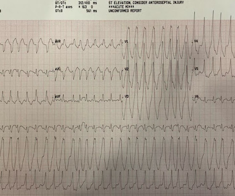
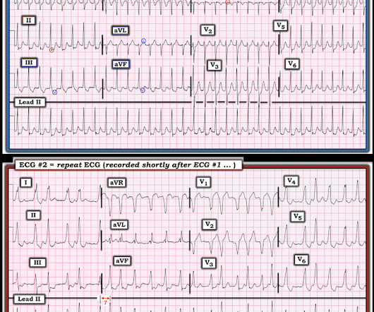
The ECG : The first impression is that is a regular WIDE COMPLEX TACHYCARDIA. The QRS duration is about 250 ms (.25 25 seconds) – VERY WIDE. It pays to take a moment to consider the possibility of REGULAR REALLY WIDE COMPLEX TACHYCARDIA (RRWCT) before making a treatment decision.

EKG/ECG 98
article thumbnail

ECG Blog #419 — The Cause of ECG #1?

Ken Grauer, MD

I was sent the 2 ECGs shown in Figure-1 — which were recorded from an elderly man whose heart beat "has been irregular for years". Regarding the 2 ECGs in Figure-1 : ECG #1 is the initial tracing obtained at the scene by the EMS ( E mergency M edical S ystems ) team — in association with an alert but markedly hypotensive patient.

EKG/ECG 471
Insiders

Sign Up for our Newsletter

This site is protected by reCAPTCHA and the Google Privacy Policy and Terms of Service apply.

Trending Sources

article thumbnail

ECG Blog #409 — Every-Other-Beat.

Ken Grauer, MD

The ECG in Figure-1 — was obtained from a patient with palpitations. Why is QRS morphology changing ? Figure-1: The initial ECG in today's case. My Interpretation of the ECG in Figure-1: Since the patient is hemodynamically stable — there is time for systematic assessment of the rhythm.

EKG/ECG 430
article thumbnail

ECG Blog #417 — AFib with Aberrancy?

Ken Grauer, MD

The ECG in Figure-1 was obtained from a previously healthy middle-aged man — who presented to the ED ( E mergency D epartment ) for shortness of breath. QUESTIONS: How would YOU interpret the ECG shown in Figure-1 ? Figure-1: The initial ECG in today's case. Figure-1: The initial ECG in today's case.

EKG/ECG 312
article thumbnail

ECG Blog #429 — Mobitz I or Mobitz II?

Ken Grauer, MD

The 12-lead ECG and long lead II rhythms shown in Figure-1 — was obtained from an older man with a recent history of “easy fatiguability” and a presyncopal episode. QUESTIONS: How would YOU interpret the ECG in Figure-1 ? Figure-1: The initial ECG in today’s case. I outline my approach for doing so below.

EKG/ECG 263
article thumbnail

ECG Blog #406 — To Do Additional Leads?

Ken Grauer, MD

For full discussion of this case — See ECG Blog #351 — == The ECG in Figure-1 — was obtained from a previously healthy older man who contacted EMS ( E mergency M edical S ervices ) because of "chest tightness" that began ~1 hour earlier. Given this history: QUESTIONS: How would YOU interpret the ECG in Figure-1 ?

EKG/ECG 375
article thumbnail

ECG Blog #422 — Was Clubbing an ECG Hint?

Ken Grauer, MD

I was sent the ECG in Figure-1 — with the following history: The patient is a young man in his early 20s — who presents to the ED ( E mergency D epartment ) because of SOB ( S hortness O f B reath ) that had been ongoing for several hours. QUESTIONS: In view of the above history — How would YOU interpret the ECG in Figure-1 ?

EKG/ECG 447