Remove ecg 15-lead-ecg
article thumbnail

ECG Blog #431 — My New ECG-Rhythm Podcasts!

Ken Grauer, MD

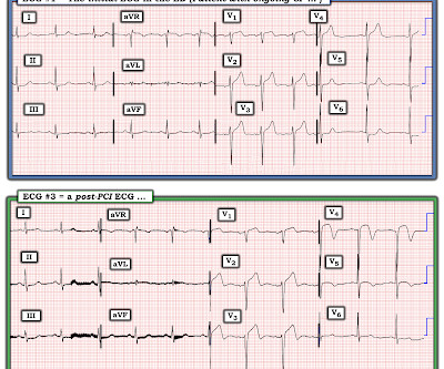
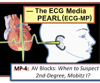
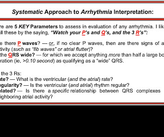
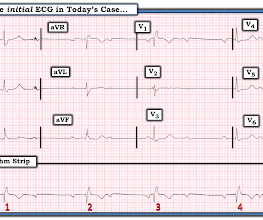
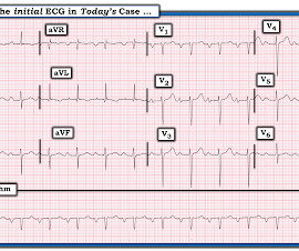
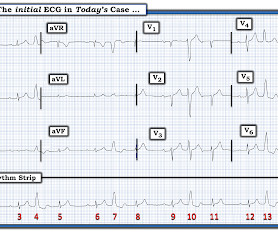
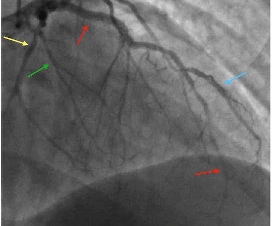
I recently recorded a series of 4 podcasts regarding KEY concepts in ECG interpretation. Easy LINKS — tinyurl.com/KG-ECG-Podcasts — [link] — Other ECG Audio PEARLS I previously made for my ECG Blog can be found in the right column of each page on this blog just below this icon — under, "ECG Audio PEARLS".

EKG/ECG 277
article thumbnail

ECG Blog #418 — A Single Lead Tells the Tale.

Ken Grauer, MD

The ECG in Figure-1 was obtained from a previously healthy man in his 40s — who presented to the ED ( E mergency D epartment ) with new-onset CP ( C hest P ain ) that awakened him from sleep. ECG #1 was recorded ~90 minutes after the patient was awakened from sleep. Does a single lead “tell the tale”? He was still having CP.

EKG/ECG 412
Insiders

Sign Up for our Newsletter

This site is protected by reCAPTCHA and the Google Privacy Policy and Terms of Service apply.

Trending Sources

article thumbnail

ECG Blog #422 — Was Clubbing an ECG Hint?

Ken Grauer, MD

I was sent the ECG in Figure-1 — with the following history: The patient is a young man in his early 20s — who presents to the ED ( E mergency D epartment ) because of SOB ( S hortness O f B reath ) that had been ongoing for several hours. QUESTIONS: In view of the above history — How would YOU interpret the ECG in Figure-1 ?

EKG/ECG 447
article thumbnail

ECG Blog #415 — The Cath showed NO Occlusion!

Ken Grauer, MD

The ECG in Figure-1 was obtained following successful resuscitation. QUESTIONS: In view of the above history — How would YOU interpret the ECG in Figure-1 ? Is this ECG finding present in today’s initial ECG? Figure-1: The initial ECG in today's case — obtained after successful resuscitation from cardiac arrest. (

EKG/ECG 393
article thumbnail

ECG Blog #417 — AFib with Aberrancy?

Ken Grauer, MD

The ECG in Figure-1 was obtained from a previously healthy middle-aged man — who presented to the ED ( E mergency D epartment ) for shortness of breath. QUESTIONS: How would YOU interpret the ECG shown in Figure-1 ? Figure-1: The initial ECG in today's case. Figure-1: The initial ECG in today's case.

EKG/ECG 312
article thumbnail

ECG Blog #396 — Why the Flat Line?

Ken Grauer, MD

The ECG in Figure-1 — was obtained from a middle-aged man with palpitations and shortness of breath. How would YOU interpret the ECG in Figure-1 ? Figure-1: The initial ECG in today's case. ( To improve visualization — I've digitized the original ECG using PMcardio ). Figure-1: The initial ECG in today's case. (

EKG/ECG 421
article thumbnail

ECG Blog #429 — Mobitz I or Mobitz II?

Ken Grauer, MD

The 12-lead ECG and long lead II rhythms shown in Figure-1 — was obtained from an older man with a recent history of “easy fatiguability” and a presyncopal episode. QUESTIONS: How would YOU interpret the ECG in Figure-1 ? Figure-1: The initial ECG in today’s case. I outline my approach for doing so below.

EKG/ECG 263