Remove ecg circumflex-occlusion
article thumbnail

Sudden shock with a Nasty looking ECG. What is it?

Dr. Smith's ECG Blog

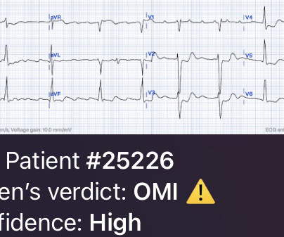
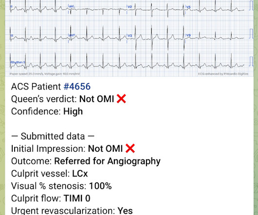
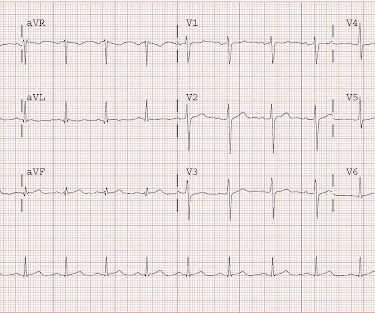
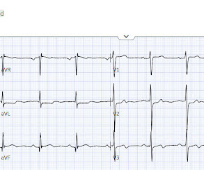
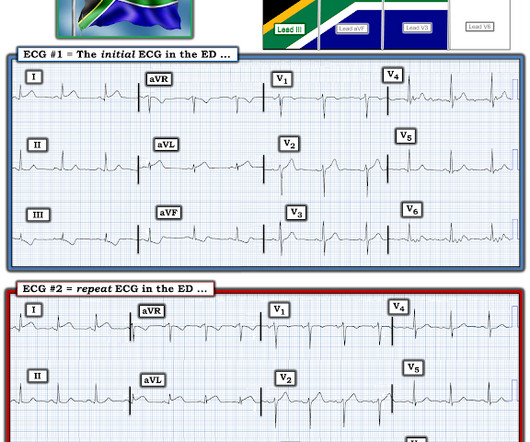
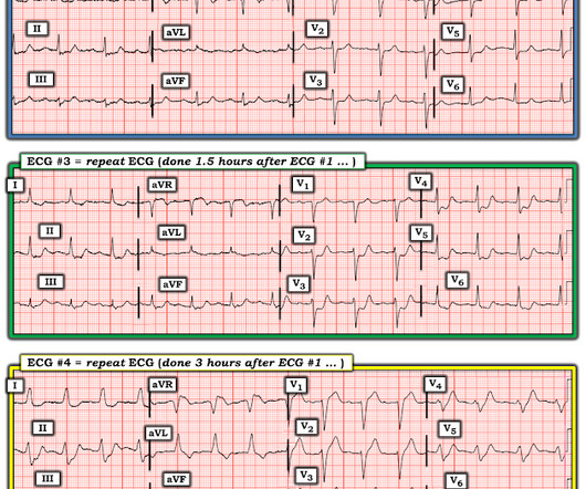
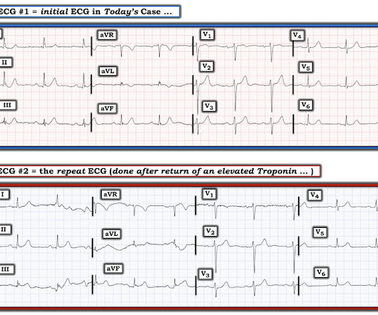
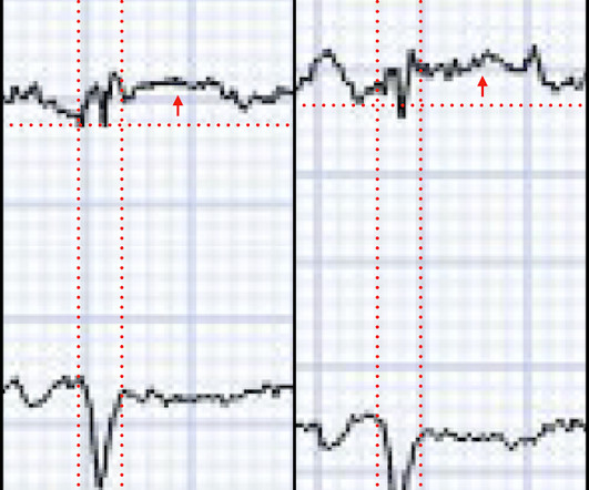
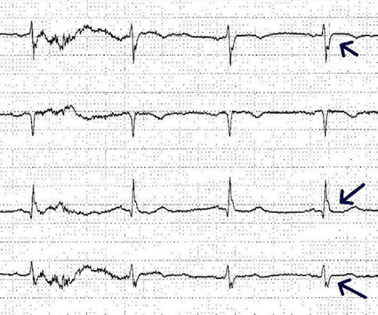
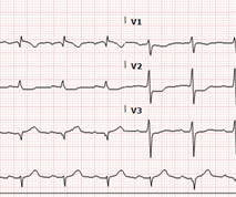
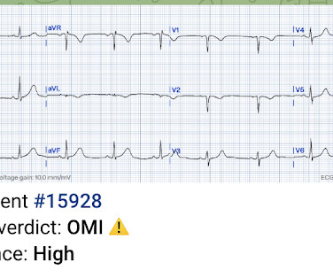
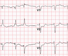
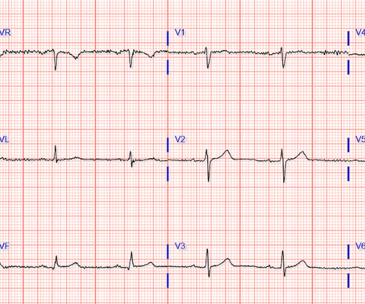
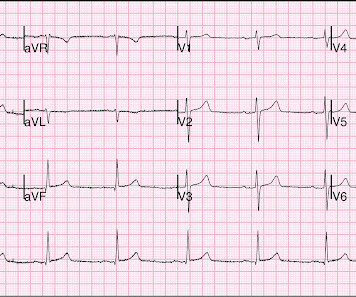
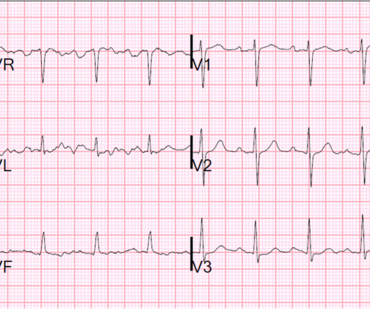
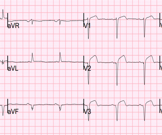
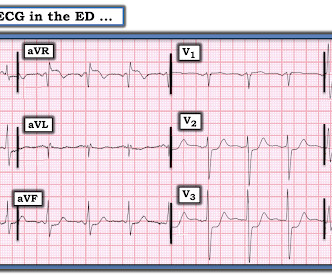
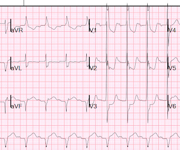
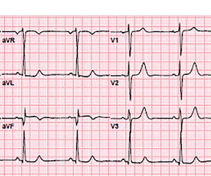
There were 2 prehospital ECGs: What do you think? When I was shown this ECG, I said it looks like such widespread ischemia that is might be a left main occlusion, or LM ischemia plus circumflex occlusion (high lateral and posterior OMI). Moreover, left main occlusion often presents near death.

Shock 98
article thumbnail

Chest pain with anterior ST depression: look what happens if you use posterior leads.

Dr. Smith's ECG Blog

Here's the prior ECG: This confirms thew anterior ST depression is new. The first ECG was labeled “anterior subendocardial ischemia”, but subendocardial ischemia does not localize. So a patient with high pretest probability (prior CABG with new chest pain), had new ECG changes showing posterior OMI. Do you need posterior leads?

EKG/ECG 100
Insiders

Sign Up for our Newsletter

This site is protected by reCAPTCHA and the Google Privacy Policy and Terms of Service apply.

Trending Sources

article thumbnail

Is there OMI on this ECG?

Dr. Smith's ECG Blog

The patient with no prior cardiac history presented in the middle of the night with acute chest pain, and had this ECG recorded during active pain: I did not see any ischemia on this electrocardiogram. I think the ECG is normal. There was a 100% circumflex occlusion that was opened. Therefore, we activate the Cath Lab.

EKG/ECG 88
article thumbnail

Infection and DKA, then sudden dyspnea while in the ED

Dr. Smith's ECG Blog

This ECG was recorded: What do you think? Another ECG was recorded: What do you think? Aslanger's is a combination of inferior OMI with widespread ST depression and is due to BOTH occlusion of one artery (usually the circumflex, but sometimes the RCA) AND simultantous 3 vessel disease. There is widespread ST depression.

EKG/ECG 88
article thumbnail

Circumflex Occlusion May be Subtle or Invisible on the ECG

Dr. Smith's ECG Blog

Case 1 Below are 2 cases of circumflex or obtuse marginal (branch of circumflex) occlusions showing how subtle they may be: A 52 y.o. Here is the EKG at 1810: A repeat ECG 1 hour later is subtly changed, with some evolving T-wave inversion best seen in V5 and V6. He reports intermittent CP and SOB for 2-3 days.

EKG/ECG 52
article thumbnail

Dueling OMI: does this 30 year old with chest pain have any signs of occlusion or reperfusion?

Dr. Smith's ECG Blog

Below is ECG #1 at triage. Are there any signs of occlusion or reperfusion? There are also subtle biphasic T waves in V3-4 of unclear significance (this can be seen in anterior or RV reperfusion, but this usually does not accompany infero-posterior occlusion). So the patient waited to be seen. What do you think?

EKG/ECG 52
article thumbnail

Occlusion myocardial infarction is a clinical diagnosis

Dr. Smith's ECG Blog

Occlusion myocardial infarction is a clinical diagnosis Written by Willy Frick (@Willyhfrick). Willy is a cardiology fellow with a keen interest in the ECG in OMI. Smith again: Whenever someone sends me an ECG for determination of OMI or Not OMI, I say: "Any patient with OMI can have an ECG that does not show OMI.

EKG/ECG 110