article thumbnail

Ultrasound in Cardiac Arrest

Mount Sinai EM

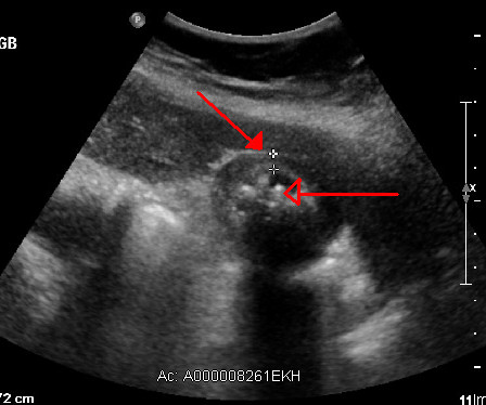
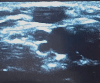
Ultrasound during cardiac arrest has quickly become standard. Initially, data suggested that the use of ultrasound during arrest increased pauses between compressions which worsens outcomes. Finally, patients with PEA and cardiac standstill on ultrasound have a 0.0%-0.6% Yours in ultrasounding, Shivam

article thumbnail

Medical Malpractice Insights: Fibroids Found on Ultrasound

EMDocs

Chuck Pilcher, MD, FACEP Editor, Medical Malpractice Insights Fibroids Found on Ultrasound Classic case of confirmation bias, appendicitis missed Facts : A middle-aged female with history of ovarian cysts and fibroids develops right lower quadrant abdominal pain rated as a 10 out of 10. To opt in to the free subscriber list, click here.

Insiders

Sign Up for our Newsletter

This site is protected by reCAPTCHA and the Google Privacy Policy and Terms of Service apply.

Trending Sources

article thumbnail

US Probe: Suprapatellar Recess Injection for Chronic Knee Pain in the Emergency Department – An Effective Approach for Relief

EMDocs

Author: Abdo Zeinoun MD, Clinical Ultrasound Fellow, Department of Emergency Medicine, Rutgers New Jersey Medical School // Reviewed by: Stephen Alerhand, MD ( @SAlerhand) ; Steve Fields, MD Patient Case A 60 year-old male with a history of hypertension presents with worsening right knee pain over the last 3 days. Why Use Ultrasound Guidance?

article thumbnail

Ultrasound Case 112

Life in the Fast Lane

Casey Parker and James Rippey Ultrasound Case 112 A 30 year old woman who is currently 30 weeks gestation presents to the ED with abrupt, severe right loin pain.

article thumbnail

US Probe: An Abundance of Artifacts

EMDocs

While performing the right upper quadrant ultrasound, you notice something that looks like the liver sitting above the diaphragm. You wonder if the patient has concomitant lower lobe pneumonia or if the ultrasound machine is broken. Why are there artifacts? What are the five assumptions made?

article thumbnail

Thoracic Aortic Emergencies on Ultrasound: Recognizing Aneurysms and Dissections

Core Ultrasound

Jailyn Avila walks you through how to diagnose thoracic aortic aneurysms and thoracic aortic dissection using bedside ultrasound. Early detection of thoracic aortic pathology can be life-saving, and ultrasound can play a key role in raising suspicion quickly.

article thumbnail

Point-of-care ultrasound for foreign bodies

Emergency Medicine Journal

A point-of-care ultrasound (POCUS) was performed to evaluate for the presence of an FB using a high frequency (6–13 MHz) linear array ultrasound (US) transducer. There is a small 2–3 mm wound inferolateral to the right knee with very mild associated erythema, no active bleeding or swelling and no palpable FB ( figure 2 ).