Remove ecg giant-t-waves
article thumbnail

ECG Blog #412 — Is Cardiac Cath Indicated?

Ken Grauer, MD

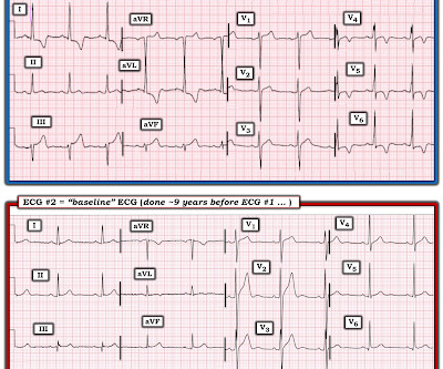
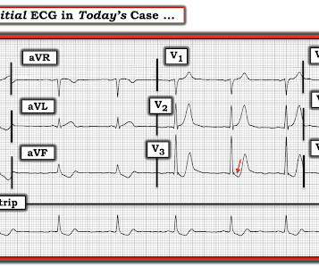
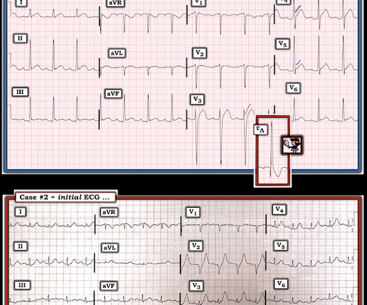
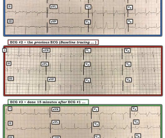
The ECG in Figure-1 was obtained from a middle-aged man with known hypertension — who presented to the ED ( E mergency D epartment ) for CP ( C hest P ain ) over the preceding 2-3 days. QUESTIONS: How would YOU interpret the ECG in Figure-1 ? The Initial ECG in Today’s CASE: The rhythm in ECG #1 is sinus at 65-70/minute.

EKG/ECG 379
article thumbnail

An elderly patient with stuttering chest pain. Don't jump to conclusions.

Dr. Smith's ECG Blog

I was reading ECGs on the system and saw this one, and instantly knew the probable ECG diagnosis: What do you think? This is a very typical ECG for Hypertrophic Cardiomyopathy. Explainability Notice it is the wide, large R-wave which catches her attention and leads to the "Not OMI" diagnosis. What do you think now?

EKG/ECG 100
Insiders

Sign Up for our Newsletter

This site is protected by reCAPTCHA and the Google Privacy Policy and Terms of Service apply.

Trending Sources

article thumbnail

What is this ECG finding? Do you understand it before you hear the clinical context?

Dr. Smith's ECG Blog

Written by Pendell Meyers First try to interpret this ECG with no clinical context: The ECG shows an irregularly irregular rhythm, therefore almost certainly atrial fibrillation. After an initially narrow QRS, there is a very large abnormal extra wave at the end of the QRS complex. C), with Cardiac Echo -- A Pathognomonic ECG.

article thumbnail

A man in his 30s with chest pain. How was he managed? What if they had used the Queen of Hearts?

Dr. Smith's ECG Blog

Triage ECG: And here she explains her assessment: The ECG was read as simply "No ST elevation." No repeat ECG was done at this time. Repeat ECG shows no changes." Here is that repeat ECG below, around 3 hours after triage: Repeat troponin during delay rose to 18,700 ng/L. Which is true. None further were ordered.

EKG/ECG 100
article thumbnail

Should we activate the cath lab? A Quiz on 5 Cases.

Dr. Smith's ECG Blog

Triage is backed up, and 10 minutes into your shift one of the ED nurses brings your several ECG s that has not been overread by a physician. ECG#1 ECG#2 ECG#3 ECG#4 ECG#5 See outcomes of all 5 below, with the Queen of Hearts AI Bot interpretation. link] ECG #1 : There is sinus rhythm, narrow QRS complexes.

article thumbnail

See this "NSTEMI" go unrecognized for what it really is, how it progresses, and what happens

Dr. Smith's ECG Blog

The baseline ECG is basically normal with no ischemia. Here is what the Queen of Hearts says about the Baseline ECG: Active chest pain triage ECG also read as not OMI: She says "not OMI", but she does not have access to the baseline ECG. In the future, we will be able to have her compare with previous and serial ECGs.

EKG/ECG 79
article thumbnail

Another myocardial wall is sacrificed at the altar of the STEMI/NonSTEMI mass delusion (and Opiate pain relief).

Dr. Smith's ECG Blog

I received the following text message with these 3 EKGs (providers text me ECGs all day every day; most are false positives; many are subtle true positives): "Hi Steve, here are 3 EKGs for you (my colleague's case). Smith Interpretation : there is diffuse ST depression followed by a hyperacute T-wave. Cath attending is aware.

EKG/ECG 70