Remove ecg left-main-coronary-artery-occlusion-0
article thumbnail

ECG Pointers: STEMI Equivalents from the American College of Cardiology

EMDocs

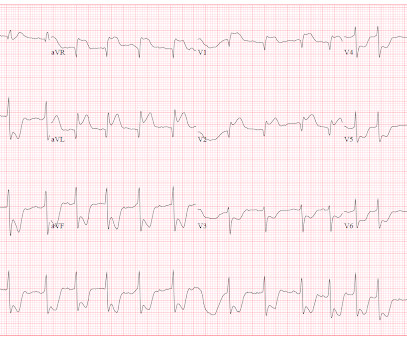
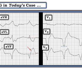
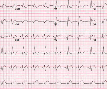
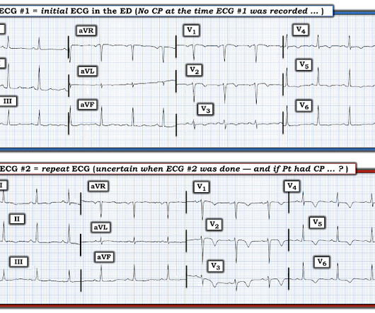
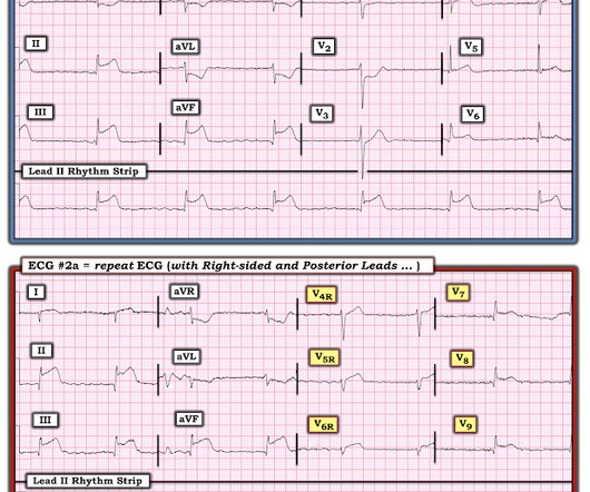
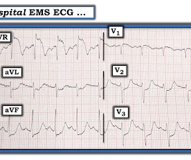
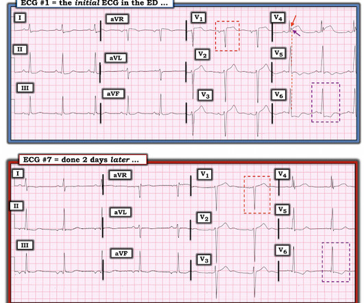
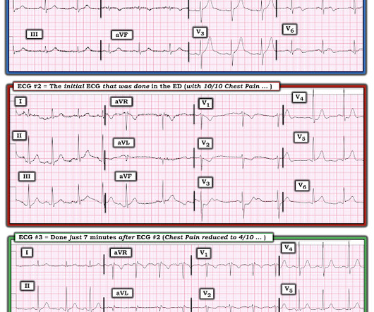
The most common diagnostic test to identify patients who might require percutaneous intervention is the electrocardiogram (ECG). Emergency physicians have recognized for some time that there are many occlusions of the coronary arteries that do not present with classic STEMI criteria on the ECG.

EKG/ECG 109
article thumbnail

How does Acute Total Left Main Coronary occlusion present on the ECG?

Dr. Smith's ECG Blog

Post by Smith and Meyers Sam Ghali ( [link] ) just asked me (Smith): "Steve, do left main coronary artery *occlusions* (actual ones with transmural ischemia) have ST Depression or ST Elevation in aVR?" Total LM occlusion can present with STE or STD in aVR. All are, however, clearly massive STEMI.

EKG/ECG 52
Insiders

Sign Up for our Newsletter

This site is protected by reCAPTCHA and the Google Privacy Policy and Terms of Service apply.

Trending Sources

article thumbnail

I'm so sorry when medics get abused for activating the cath lab

Dr. Smith's ECG Blog

Cortland Ashbrook from Spokane County, Washington, sent this message: Hey doctor Smith, I wondered if you’d give me your opinion on these ECG tracings I took as a paramedic in the field? Case: The call was an elderly gentleman who was at home when he experienced a sudden onset of vague chest discomfort along with nausea, and left arm aching.

EKG/ECG 52
article thumbnail

A teenager with chest pain, a troponin below the limit of detection, and "benign early repolarization"

Dr. Smith's ECG Blog

Meyers ECG Interpretation: Easily diagnostic of LAD occlusion. The ECG easily meets STEMI criteria in all leads V2-V6, as well. I sent this ECG to Dr. Smith, with the only information that it is a 17 year old with chest pain. The ECG was interpreted as "benign early repolarization."

EKG/ECG 52
article thumbnail

A 30-something woman with intermittent CP, a HEART score of 2 and a Negative CT Coronary Angiogram on the same day

Dr. Smith's ECG Blog

I do not have her previous ECGs, but reportedly these T-wave inversions were not present previously. However, when there is evolution of the T-waves, as with the 2nd ECG, the specificity for Wellens' is much higher. A CT Coronary angiogram was ordered. Here are the results: --Minimally obstructive coronary artery disease. --LAD

EKG/ECG 52
article thumbnail

Chest pain and shock: Is there a right ventricular OMI on this ECG? And should he undergo trancutaneous pacing?

Dr. Smith's ECG Blog

His prehospital ECG was diagnostic of inferior posterior OMI. Here is his ED ECG: There is bradycardia with a junctional escape. We recorded an ECG in which V1-V3 were put in the position of V4R-V6R, and V4-6 were placed in V7-9 to (academically) confirm posterior OMI. Not all proximal RCA occlusion results in RV MI (i.e.,

Shock 71
article thumbnail

What do you think the echocardiogram shows in this case?

Dr. Smith's ECG Blog

Here is the EMS ECG: Obviously massive diffuse subendocardial ischemia, with profound STD and STE in aVR Of course this pattern is most often seen from etoliogies other than ACS. The ECG only tells you there is ischemia, not the etiology of it. Angiogram: Severe diffuse left main disease, up to 80% at the ostial left main.

EKG/ECG 52