Remove ecg premature-ventricular-contraction
article thumbnail

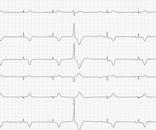
Sinus Bradycardia and More

ECG Guru

Let's analyze the ECG. This is followed by a premature ventricular contraction (PVC). This cannot be conducted to the ventricles, either because the ventricular myocardium is still unexcitable or the PVC has conducted retrogradely into the AV node and this is therefore still refractory.

EKG/ECG 52
article thumbnail

Physical Examination as a Helpful Aid in Decision-Making in Challenging ECGs

Dr. Smith's ECG Blog

An initial electrocardiogram (ECG) is provided below. A subsequent ECG was taken: There is persistent ST elevation, especially being concordant in inferolateral leads. While the initial impression might not immediately suggest ventricular tachycardia (VT), a closer examination raises suspicion. What do you think?

EKG/ECG 84
Insiders

Sign Up for our Newsletter

This site is protected by reCAPTCHA and the Google Privacy Policy and Terms of Service apply.

Trending Sources

article thumbnail

Orthostatic hypotension onset after invasive procedure?

Dr. Smith's ECG Blog

The next morning, the following ECG was obtained. Ventricular lead This tracing may also show failure to sense in the ventricular lead. The ventricular lead should have sensed a native QRS complex and suppressed pacing, but it did not, and therefore paced inappropriately. Repeat ECG from the emergency department is shown.

EKG/ECG 88
article thumbnail

MI in Children

Pediatric Emergency Playbook

With every systolic contraction, the artery is occluded. Electrocardiography (ECG) should be performed on any patient with significant blunt chest injury. A negative ECG is highly consistent with no significant blunt myocardial injury. A solitary elevated troponin without ECG abnormality is of unclear significance.