Remove ecg lewis-lead
article thumbnail

ECG Blog #425 — Are there P Waves?

Ken Grauer, MD

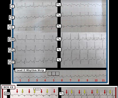
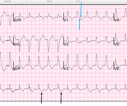
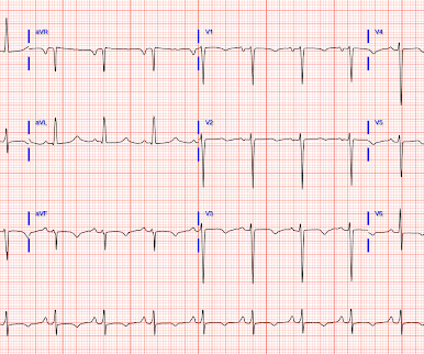
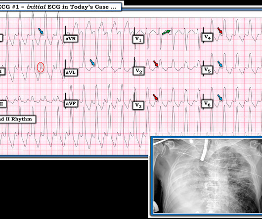
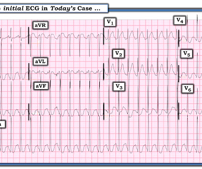
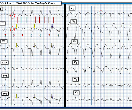
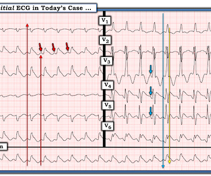
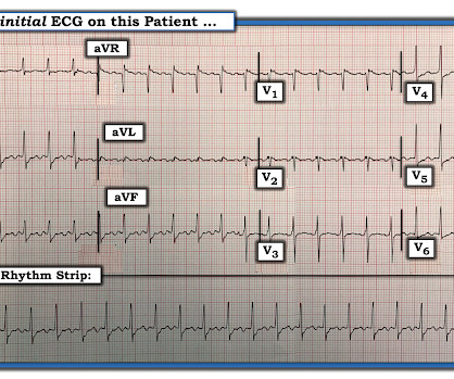
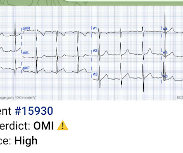
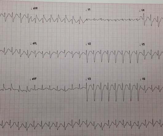
I was sent the ECG in Figure-1 — told only that the patient was 70 years old, and had a history of an ASD ( A trial S eptal D efect ). The patient was hemodynamically stable with ECG #1. Figure-1: The initial ECG in today's case. Serum K+ was normal. QUESTIONS: How would YOU interpret the rhythm in Figure-1 ? Are there P waves?

EKG/ECG 284
article thumbnail

Wide complex tachycardia, resistant to cardioversion. Some fascinating features here.

Dr. Smith's ECG Blog

Here is her ECG: Regular Wide Complex Tachycardia. She had an ECG done yesterday!! So we applied Lewis Leads. How do we place Lewis Leads? -- Use the monitor leads (not the 12-lead ECG): 1. Monitor Lead I. It’s a very interesting ECG I saw yesterday. What do you think?

EKG/ECG 104
Insiders

Sign Up for our Newsletter

This site is protected by reCAPTCHA and the Google Privacy Policy and Terms of Service apply.

Trending Sources

article thumbnail

How will you save this critically ill patient? A fundamental and lifesaving ECG interpretation that everyone must recognize instantly.

Dr. Smith's ECG Blog

An ECG was performed: What do you think? It is unclear what changes happened to the rhythm based on the EMS interventions, but the patient arrived to the ED remaining critically ill and with a very wide complex reported (no ECGs from ED available sadly). Here is her ECG the next day with normal potassium level: She did well.

EKG/ECG 123
article thumbnail

VT? Or Supraventricular tachycardic rhythm with aberrancy?

Dr. Smith's ECG Blog

The following 12-lead was obtained: What do you think? There are possible P waves in lead II. Certainty could come with the use of Lewis leads. Here is a normal LBBB: In LBBB, monophasic wide R-waves should be limited to the lateral leads in left bundle branch block, as in this ECG.

EKG/ECG 124
article thumbnail

A young man with tachycardia. Should We Try Adenosine?

Dr. Smith's ECG Blog

An ECG was recorded: What do you think? You must look closely for P-waves, and if you do, the P-waves are obvious (if you look at the leads most likely to reveal P-waves: leads II and V1). In lead II, the P-wave is always upright and has the greatest amplitude because the atrium depolarizes towards the apex (lead II).

EKG/ECG 78
article thumbnail

Four patients with chest pain and ‘normal’ ECG: can you trust the computer interpretation?

Dr. Smith's ECG Blog

All initial ECGs were labeled ‘normal’ or ‘otherwise normal’ by the computer interpretation, and below are the ECGs with the final cardiology interpretation. 1-3] But these studies were very short duration and used cardiology interpretation of ECGs or emergent angiography rather than patient outcomes.

EKG/ECG 108
article thumbnail

A fascinating electrophysiology case. What is this wide complex tachycardia, and how best to manage it?

Dr. Smith's ECG Blog

The ECG below was recorded. The ECG was interpreted as showing atrial flutter with 2:1 conduction. Answer : The ECG above shows a regular wide complex tachycardia. Also, lead I could give the initial impression of showing flutter waves. How do you interpret the below ECG? What is your assessment?

EKG/ECG 86