Remove ecg sinus-block
article thumbnail

ECG Blog #421 — Has there been a Recent MI?

Ken Grauer, MD

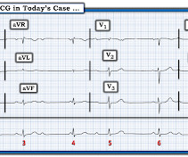
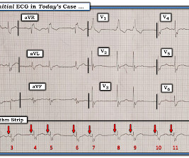
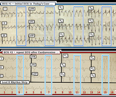
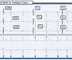
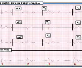
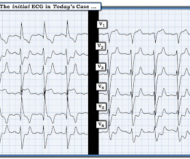
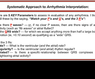
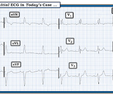
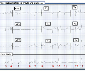
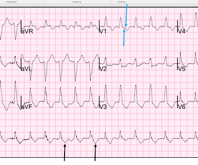
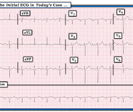
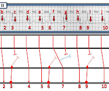
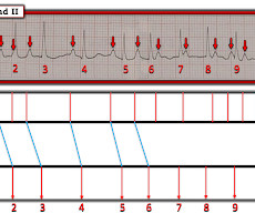
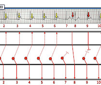
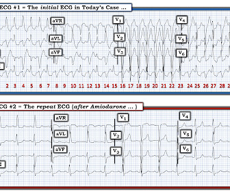
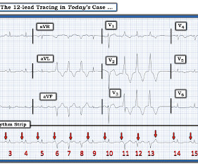
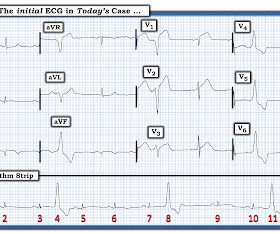
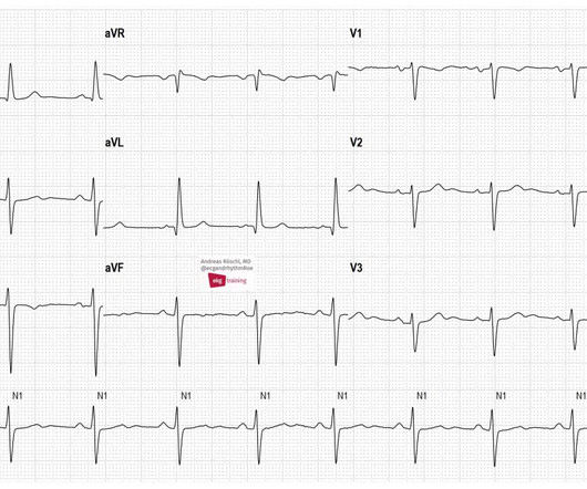
What if you were asked to interpret the ECG in Figure-1 ? Figure-1: The initial ECG in today's case. ( To improve visualization — I've digitized the original ECG using PMcardio ). By the P s, Q s, 3 R Approach ( which I review in ECG Blog #185 ): Lots of P waves are present — being well seen in the long lead II rhythm strip.

EKG/ECG 390
article thumbnail

ECG Blog #419 — The Cause of ECG #1?

Ken Grauer, MD

I was sent the 2 ECGs shown in Figure-1 — which were recorded from an elderly man whose heart beat "has been irregular for years". Regarding the 2 ECGs in Figure-1 : ECG #1 is the initial tracing obtained at the scene by the EMS ( E mergency M edical S ystems ) team — in association with an alert but markedly hypotensive patient.

EKG/ECG 471
Insiders

Sign Up for our Newsletter

This site is protected by reCAPTCHA and the Google Privacy Policy and Terms of Service apply.

Trending Sources

article thumbnail

ECG Blog #429 — Mobitz I or Mobitz II?

Ken Grauer, MD

The 12-lead ECG and long lead II rhythms shown in Figure-1 — was obtained from an older man with a recent history of “easy fatiguability” and a presyncopal episode. QUESTIONS: How would YOU interpret the ECG in Figure-1 ? Is the group beating due to the Mobitz I or Mobitz II type of 2nd-degree AV block?

EKG/ECG 263
article thumbnail

ECG Blog #405 — Is AV Block Complete (vs AV Dissociation)

Ken Grauer, MD

For full discussion of this case — See ECG Blog #191 — == The 2-lead rhythm strip shown in Figure-1 was obtained from an elderly woman who presented to the ED following a syncopal episode. On the basis of this rhythm strip — she was diagnosed as being in complete AV Block. Figure-1: Is this complete AV Block ? ( 19:40 min.

EKG/ECG 421
article thumbnail

ECG Blog #422 — Was Clubbing an ECG Hint?

Ken Grauer, MD

I was sent the ECG in Figure-1 — with the following history: The patient is a young man in his early 20s — who presents to the ED ( E mergency D epartment ) because of SOB ( S hortness O f B reath ) that had been ongoing for several hours. QUESTIONS: In view of the above history — How would YOU interpret the ECG in Figure-1 ?

EKG/ECG 447
article thumbnail

ECG Blog #411 — Is it Wenckebach?

Ken Grauer, MD

Figure-1: The initial ECG in today's case. KEY Clinical Point: If I was the medical provider charged with the care of the patient whose ECG is shown in Figure-1 — I would approach this tracing in the following sequential stages: I’d first establish that the patient was hemodnamically stable with this ECG and this cardiac rhythm.

EKG/ECG 417
article thumbnail

ECG Blog #414 — What Kind of AV Block?

Ken Grauer, MD

I was sent the ECG in Figure-1 — without the benefit of any history. QUESTION: What kind of AV block is present in Figure-1 ? Figure-1: The initial ECG in today's case. ( To improve visualization — I've digitized the original ECG using PMcardio ). The Q RS complex is narrow in all 12 leads. The rhythm is not R egular.

EKG/ECG 255