Remove ecg spontaneous-reperfusion
article thumbnail

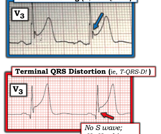
ECG Blog #426 — Are STEMI Criteria Met?

Ken Grauer, MD

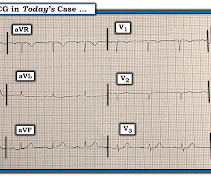
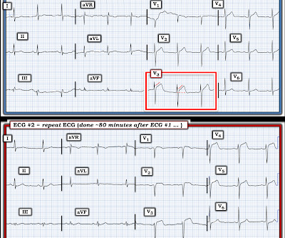
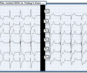
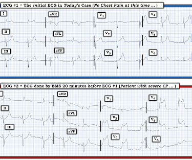
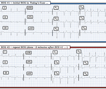
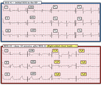
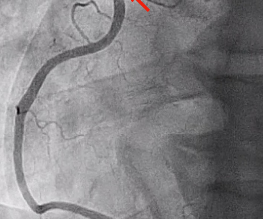
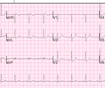
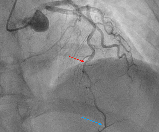
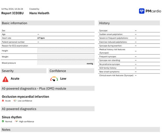
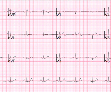
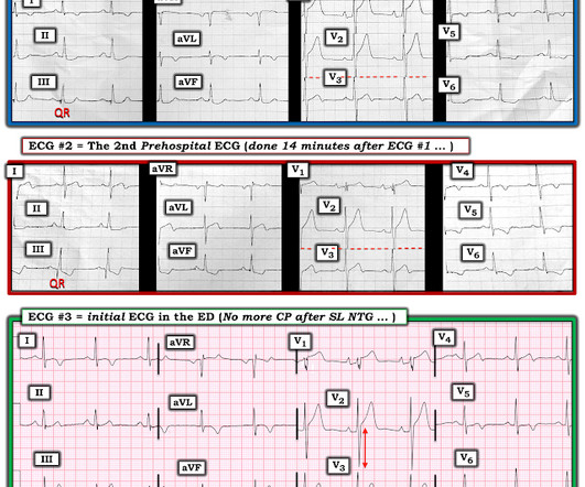
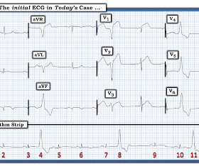
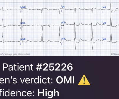
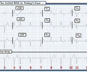
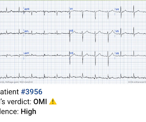
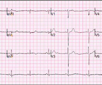
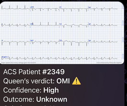
The ECG in Figure-1 — was obtained from a 70-ish year old man with episodic CP ( C hest P ain ) over the previous 2-3 days , being awakened from sleep now for a more severe CP episode. QUESTIONS: In view of this history — How would YOU interpret this ECG? Figure-1: The initial ECG in today's case.

EKG/ECG 396
article thumbnail

ECG Blog #418 — A Single Lead Tells the Tale.

Ken Grauer, MD

The ECG in Figure-1 was obtained from a previously healthy man in his 40s — who presented to the ED ( E mergency D epartment ) with new-onset CP ( C hest P ain ) that awakened him from sleep. ECG #1 was recorded ~90 minutes after the patient was awakened from sleep. Figure-1: The initial ECG in today's case. (

EKG/ECG 412
Insiders

Sign Up for our Newsletter

This site is protected by reCAPTCHA and the Google Privacy Policy and Terms of Service apply.

Trending Sources

article thumbnail

ECG Blog #430 — Just a Regular LBBB ECG?

Ken Grauer, MD

The ECG in Figure-1 — was obtained from an older man who had just completed dialysis — and , is now complaining of abdominal discomfort that radiates to his chest. Figure-1: The initial ECG in today's case. ( To improve visualization — I've digitized the original ECG using PMcardio ). How would YOU approach this case?

EKG/ECG 374
article thumbnail

ECG Blog #392 — Repolarization T Waves?

Ken Grauer, MD

The ECG in Figure-1 was obtained from a man in his 60s — who described the sudden onset of "chest tightness" that began 20 minutes earlier, but who now ( at the time this ECG was recorded ) — was no longer having symptoms. In view of this history — How would YOU interpret this ECG? Figure-1: The initial ECG in today's case. (

EKG/ECG 342
article thumbnail

ECG Blog #406 — To Do Additional Leads?

Ken Grauer, MD

For full discussion of this case — See ECG Blog #351 — == The ECG in Figure-1 — was obtained from a previously healthy older man who contacted EMS ( E mergency M edical S ervices ) because of "chest tightness" that began ~1 hour earlier. Given this history: QUESTIONS: How would YOU interpret the ECG in Figure-1 ?

EKG/ECG 375
article thumbnail

ECG Blog #415 — The Cath showed NO Occlusion!

Ken Grauer, MD

The ECG in Figure-1 was obtained following successful resuscitation. QUESTIONS: In view of the above history — How would YOU interpret the ECG in Figure-1 ? Is this ECG finding present in today’s initial ECG? Figure-1: The initial ECG in today's case — obtained after successful resuscitation from cardiac arrest. (

EKG/ECG 393
article thumbnail

ECG Blog #400 — Is this a NSTEMI?

Ken Grauer, MD

The ECG in Figure-1 is from an older man with known coronary disease — who presents to the ED ( E mergency D epartment ) with new CP ( C hest P ain ) over the past several days. QUESTIONS: In view of this history — How would you interpret the ECG in Figure-1 ? Figure-1: The initial ECG in today's case. Troponin is pending.

EKG/ECG 239