Remove ecg anti-tachycardia
article thumbnail

REBEL Core Cast 109.0 – Na Channel Blocker Poisoning

RebelEM

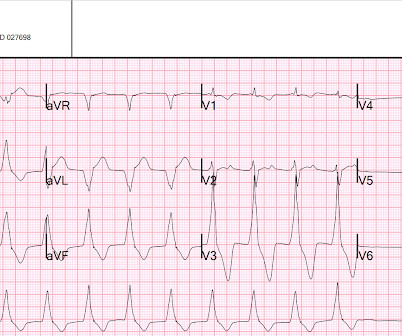
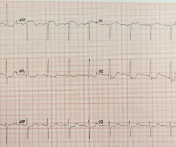
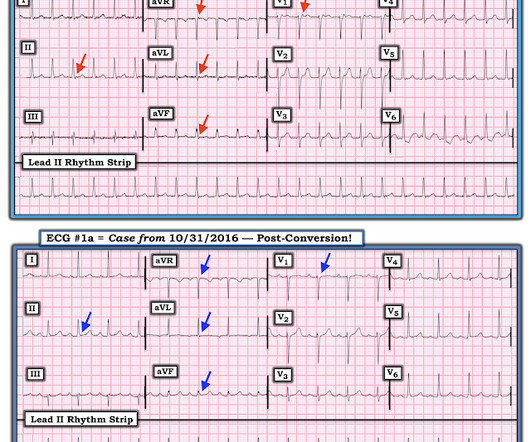
A newly “wide QRS”, especially with hemodynamic instability, should prompt consideration of sodium channel blockade and not ventricular tachycardia. Authors evaluated the relationship between QRS duration and negative clinical events in patients with confirmed tricyclic anti-depressant (TCA) poisoning. Boehnert 1985 ). Read more

article thumbnail

Young man with Gunshot wound to right chest with hemorrhagic shock, but bullet path not near heart

Dr. Smith's ECG Blog

There were times when it would be usurped by sinus tachycardia, then return to this rhythm. AIVR should never be treated with anti-dyrrhythmics!! Figure-1: The ECG sent to Ken Grauer ( showing some semblance of "group" beating ). CT of chest showed the bullet path through his right lung but nowhere near his heart.

Shock 99
Insiders

Sign Up for our Newsletter

This site is protected by reCAPTCHA and the Google Privacy Policy and Terms of Service apply.

Trending Sources

article thumbnail

Cardiac Rhythms/ECG Module

Don't Forget the Bubbles

Add into this that the majority of children will be in normal sinus rhythm (NSR) by the time of assessment so to truly identify those who have something wrong we have to be confident in identifying arrhythmias where they are present and critical when analysing an ECG in NSR. All were examined and 98% had an ECG.

EKG/ECG 98
article thumbnail

Diabetic Ketoacidosis in Paediatrics

Mind The Bleep

ECG: to monitor T wave changes due to hypokalaemia. ECG features of Hypokalaemia: Increased P wave amplitude (peaked P waves) Prolonged PR interval Widespread ST depression T wave flattening or inversion Prominent U waves (most noticeable in the precordial leads) Figure 2 : ECG of a patient with serum K+ of 1.9

article thumbnail

EM@3AM: Cardiac Transplant Complications

EMDocs

His electrocardiogram (ECG) is shown below: His chest x-ray is shown below: The patient’s high sensitivity troponin I is elevated to 3,000, and his Brain-Natriuretic Peptide (BNP) is elevated to 14,000 ng/L. 5 Treatment of supraventricular tachycardia (SVT): Therapies such as vagal maneuvers or atropine are not usually effective.

EMS 63
article thumbnail

90 year old with acute chest and epigastric pain, and diffuse ST depression with reciprocal STE in aVR: activate the cath lab?

Dr. Smith's ECG Blog

What is your ECG interpretation and what would you do next? This ECG shows a normal sinus rhythm with a normal conduction pattern (normal PR, normal QRS, and normal QTc), normal axis, late R wave progression (and misplaced V2), normal voltages, ST-elevation in aVR and global ST-depressions. BP was 110 and oxygen saturation was normal.

EKG/ECG 52
article thumbnail

New Onset Heart Failure and Frequent Prolonged SVT. What is it? Management?

Dr. Smith's ECG Blog

Here was his ED ECG: There is sinus tachycardia (rate about 114) with nonspecific ST-T abnormalities. An ECG was recorded: This shows a regular narrow complex tachycardia at a rate of about 160. See my quick review of atrial tachycardia below) The tachycardia spontaneously resolved. C (99 °F), Resp (!)

EKG/ECG 52