Remove ecg lbbb
article thumbnail

ECG Blog #430 — Just a Regular LBBB ECG?

Ken Grauer, MD

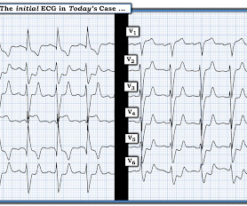
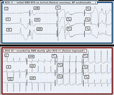
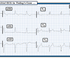
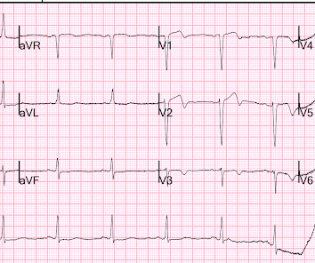
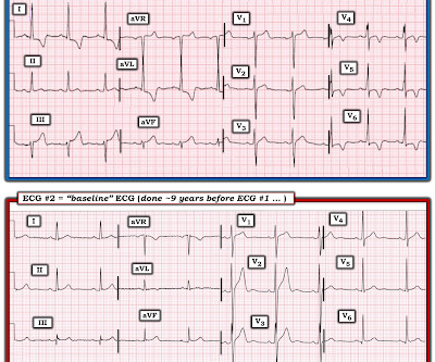
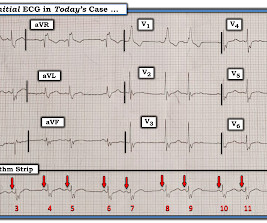
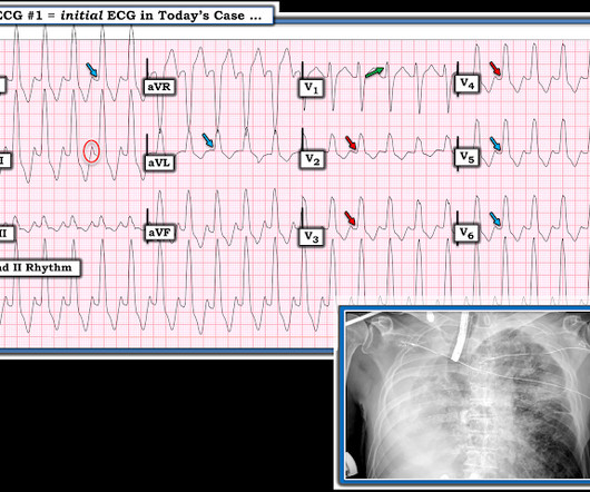
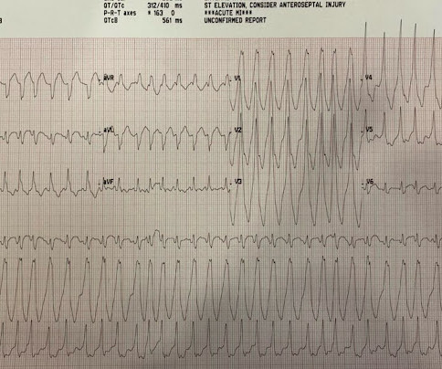
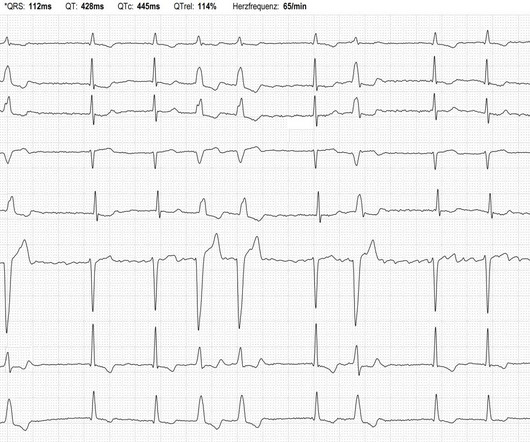
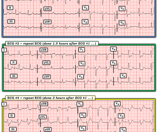
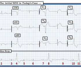
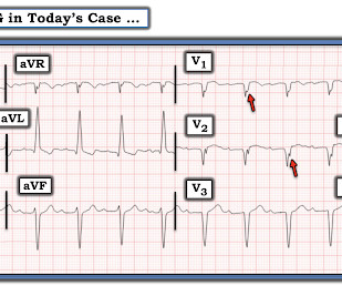
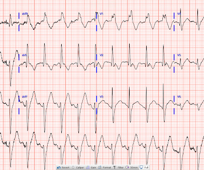
The ECG in Figure-1 — was obtained from an older man who had just completed dialysis — and , is now complaining of abdominal discomfort that radiates to his chest. The consultant interpreted this tracing as “LBBB” ( L eft B undle B ranch B lock ) — but not indicative of anything acute. Figure-1: The initial ECG in today's case. (

EKG/ECG 374
article thumbnail

ECG Blog #419 — The Cause of ECG #1?

Ken Grauer, MD

I was sent the 2 ECGs shown in Figure-1 — which were recorded from an elderly man whose heart beat "has been irregular for years". Regarding the 2 ECGs in Figure-1 : ECG #1 is the initial tracing obtained at the scene by the EMS ( E mergency M edical S ystems ) team — in association with an alert but markedly hypotensive patient.

EKG/ECG 471
Insiders

Sign Up for our Newsletter

This site is protected by reCAPTCHA and the Google Privacy Policy and Terms of Service apply.

Trending Sources

article thumbnail

ECG Blog #394 — Is QRS Morphology Disguised?

Ken Grauer, MD

The ECG in Figure-1 — was obtained from an older man with a history of prior infarction and coronary bypass surgery. Figure-1: The ECG in today's case. ( To improve visualization — I've digitized the original ECG using PMcardio ). There is no ECG indication of chamber enlargement — ST-T wave changes do not look acute.

EKG/ECG 284
article thumbnail

ECG Video Blog #403 (220) — Ps,Qs,3Rs Approach to this Tachycardia.

Ken Grauer, MD

For full discussion of this case — See ECG Blog #220 — == The long lead II rhythm strip shown in Figure-1 was obtained from an 51-year-old man who presented to the ED ( Emergency Department ) with "palpitations" that began 1 hour earlier. Figure: 12-lead ECG obtained from an 51-year-old man with palpitations.

EKG/ECG 456
article thumbnail

Explain this ECG in the context of active chest pain, slightly elevated troponin without a delta, RCA culprit, and previous with LBBB

Dr. Smith's ECG Blog

Time 17 minutes Not much different One month earlier This is Left Bundle Branch Block (LBBB) without any sign of ischemia. It turns out that she spends much of her time in LBBB (see ECGs below) What is going on??? LBBB resolves and there is V1-V3 T-wave inversion. What explains the T-wave inversions in V1-V3?

EKG/ECG 74
article thumbnail

ECG Cases 11: LBBB + Occlusion MI

Emergency Medicine Cases

In ECG Cases 11 - LBBB + Occlusion MI Jesse McLaren runs through 5 patients who present with potentially ischemic symptoms and LBBB and explains the evolution of decision making for cath lab activation criteria. The post ECG Cases 11: LBBB + Occlusion MI appeared first on Emergency Medicine Cases.

EKG/ECG 52
article thumbnail

ECG Blog #406 — To Do Additional Leads?

Ken Grauer, MD

For full discussion of this case — See ECG Blog #351 — == The ECG in Figure-1 — was obtained from a previously healthy older man who contacted EMS ( E mergency M edical S ervices ) because of "chest tightness" that began ~1 hour earlier. Given this history: QUESTIONS: How would YOU interpret the ECG in Figure-1 ?

EKG/ECG 375