


Non-respiratory Sinus Arrhythmia
ECG Guru
JULY 26, 2023
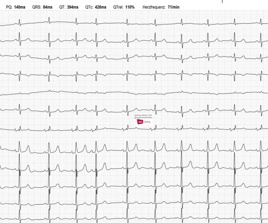
This is the ECG of an 81-year-old man with hypertension. There is a non-respiratory sinus arrhythmia present, which is essentially the minimal variant of a sick sinus syndrome. Currently, he has no complaints: no palpitations, no shortness of breath, no syncope, no chest pain.


Let's personalize your content