Remove ecg torsades-de-pointes
article thumbnail

ECG Pointers: Recurrent and Refractory Torsades de Pointes

EMDocs

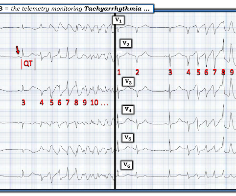
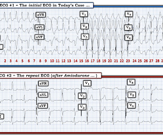
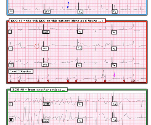
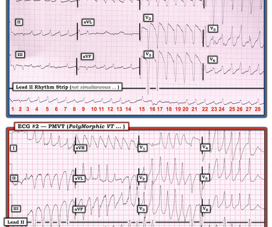
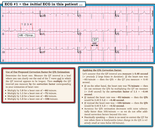
An ECG is performed and is shown below: Figure 1. Another ECG is obtained and shown below. Source: [link] As you are calling the ICU and cardiology team, the patient has recurrence of her symptoms and repeat ECG shows return of the PVT. Although commonly referred to as torsades de pointes (TdP), not all PVT is TdP.

EKG/ECG 71
article thumbnail

A 20-something woman with cardiac arrest.

Dr. Smith's ECG Blog

A few days into her hospital stay she developed chest discomfort and the following ECG was recorded. Are these ECG changes related to the CNS infection perhaps? When I saw the ECG of this patient I saw that there was definitely something "off". I sent the top ECG to Dr Smith with no information and without the baseline ECG.

EKG/ECG 90
Insiders

Sign Up for our Newsletter

This site is protected by reCAPTCHA and the Google Privacy Policy and Terms of Service apply.

Trending Sources

article thumbnail

Does this T wave pattern mean anything?

Dr. Smith's ECG Blog

An Initial ECG was performed: Initial ECG: Sinus tachycardia with prolonged QT interval (QTc of 534 ms by Bazett). A repeat ECG was performed 2 hours after arrival: QTc prolongation ato 722 ms now with alternating T wave pattern (T wave alternans) I texted this to Smith who responded: “T wave alternans and long QT. 2017.3191.

EKG/ECG 105
article thumbnail

A man in his 60s with syncope and ST depression. What does the ECG mean?

Dr. Smith's ECG Blog

Here is his triage ECG: What do you think? What is the most likely cause of the patient’s ECG findings, and what would be your first step in management? What does the ECG show? What does the ECG show? Trostel and Meyers brings home a number of important points regarding assessment of the QT interval. Is it STEMI?

EKG/ECG 97
article thumbnail

ECG Blog #363 — How Many Directions?

Ken Grauer, MD

The ECG in Figure-1 was obtained from an older woman — who presented with chest pain and palpitations over the previous hour. Figure-1: ECG obtained from an older woman with palpitations. To improve visualization — I've digitized the original ECG using PMcardio ). BP = 140/90 mm Hg in association with the rhythm in Figure-1.

EKG/ECG 195
article thumbnail

Every second counts: giant ECG abnormality after every second beat

Emergency Medicine Journal

After successful basic life support and stabilized circulation, the following ECG was recorded (see figure 1 ). Question What is the possible explanation for these ECG features? artefact on ECG bigemin premature ventricular beats.( artefact on ECG bigemin premature ventricular beats.( figure 1 )? figure 1 )?

article thumbnail

Cardiac Arrest. What does the ECG show? Also see the bizarre Bigeminy.

Dr. Smith's ECG Blog

I was texted this ECG in real time, but it turns out to actually be the 2nd one recorded in the ED. The go to the left and find that same point under aVR/aVL/aVF and under I, II, III, and you can draw this line back up to find the end of the QRS in these leads. These are frequently found in cases of Torsades. What do you think?

EKG/ECG 52