Remove ecg v4-right
article thumbnail

ECG Blog #430 — Just a Regular LBBB ECG?

Ken Grauer, MD

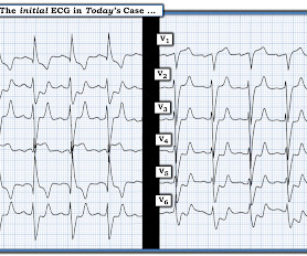
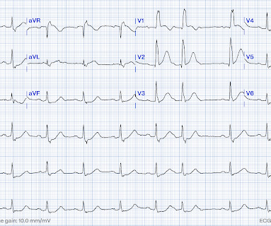
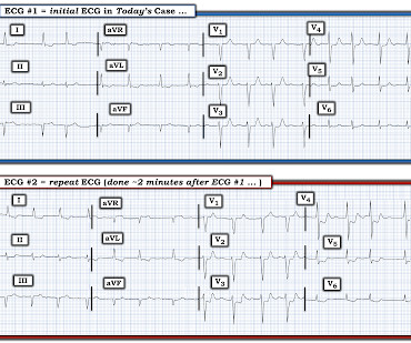
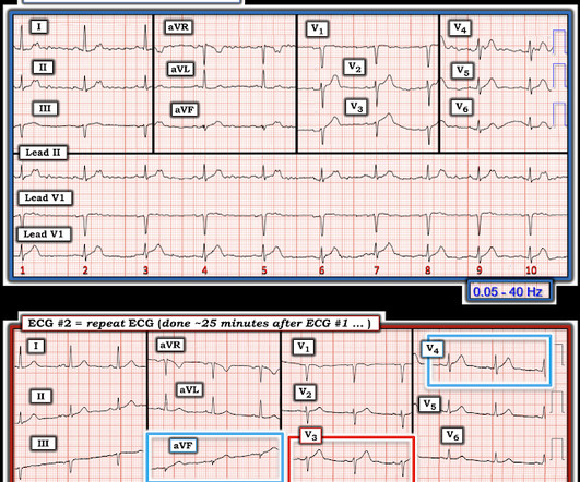
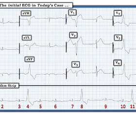
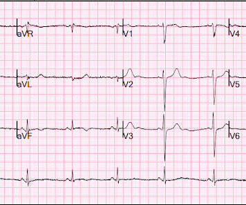
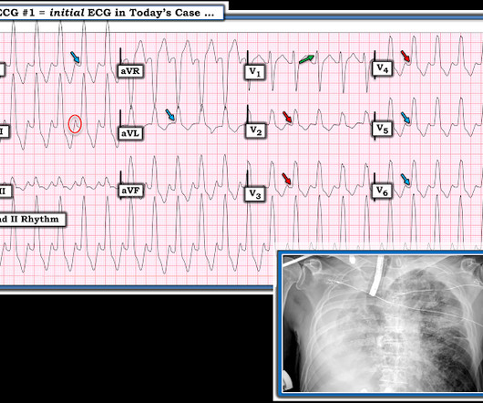
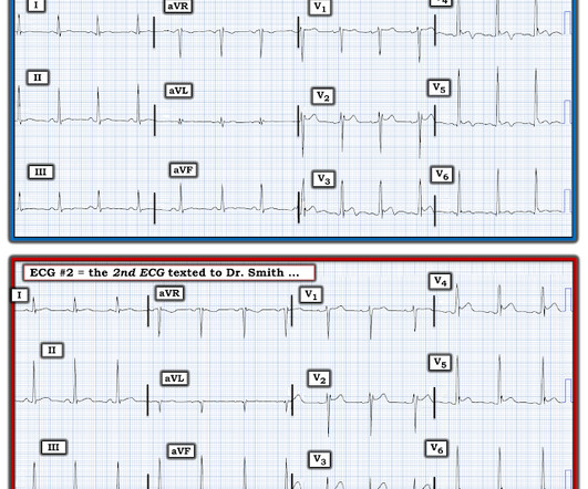
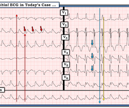
The ECG in Figure-1 — was obtained from an older man who had just completed dialysis — and , is now complaining of abdominal discomfort that radiates to his chest. Figure-1: The initial ECG in today's case. ( To improve visualization — I've digitized the original ECG using PMcardio ). How would YOU approach this case?

EKG/ECG 374
article thumbnail

ECG Blog #410 — How Tall are the T Waves?

Ken Grauer, MD

QUESTION: HOW would YOU interpret the ECG in Figure-1 — if no clinical information was provided? Figure-1: The initial ECG in today's case. ( To improve visualization — I've digitized the original ECG using PMcardio ). = The ECG in Figure-1 — was obtained following successful resuscitation.

EKG/ECG 388
Insiders

Sign Up for our Newsletter

This site is protected by reCAPTCHA and the Google Privacy Policy and Terms of Service apply.

Trending Sources

article thumbnail

ECG Blog #409 — Every-Other-Beat.

Ken Grauer, MD

The ECG in Figure-1 — was obtained from a patient with palpitations. Figure-1: The initial ECG in today's case. My Interpretation of the ECG in Figure-1: Since the patient is hemodynamically stable — there is time for systematic assessment of the rhythm. Figure-2: I've labeled the initial ECG in today's case. Figure-2 ).

EKG/ECG 430
article thumbnail

ECG Blog #406 — To Do Additional Leads?

Ken Grauer, MD

For full discussion of this case — See ECG Blog #351 — == The ECG in Figure-1 — was obtained from a previously healthy older man who contacted EMS ( E mergency M edical S ervices ) because of "chest tightness" that began ~1 hour earlier. Given this history: QUESTIONS: How would YOU interpret the ECG in Figure-1 ?

EKG/ECG 375
article thumbnail

ECG Blog #380 — What is "Swirl"?

Ken Grauer, MD

The ECG in Figure-1 — was obtained from an older woman with persistent CP ( C hest P ain ) over the previous day. Figure-1: The initial ECG in today's case. Voltage for LVH is satisfied — at least by Peguero Criteria ( Sum of deepest S in any chest lead + S in V4 ≥23 mm in a woman — as discussed in ECG Blog #73 ).

EKG/ECG 342
article thumbnail

ECG Blog #388 — Why Does Lead V1 Look Funny?

Ken Grauer, MD

The ECG in Figure-1 was obtained from an 18-year old woman — who moments before been resuscitated from out-of-hospital cardiac arrest. How would YOU interpret her post-resuscitation ECG? Does this ECG in Figure-1 provide clue(s) to the etiology of this patient's cardiac arrest? QUESTIONS: In light of the above clinical history.

EKG/ECG 369
article thumbnail

ECG Blog #369 — 10 Minutes Later.

Ken Grauer, MD

The ECG in Figure-1 was obtained from a man in his mid-60s — who presented with new chest pain. Figure-1: T he initial ECG in today’s case. MY Thoughts on the Initial ECG: The rhythm in ECG #1 — is sinus at ~70/minute. Figure-2: I've labeled significant ST-T wave findings in the limb leads of ECG #1.

EKG/ECG 195