Remove transvenous-pacemaker
article thumbnail

EM Quick Hits 20 Imaging Renal Colic, Human Trafficking, Atrial Fibrillation During COVID, Transvenous Pacemaker Placement, COVID Lung POCUS, COVID Derm, Virtual Simulation

Emergency Medicine Cases

The post EM Quick Hits 20 Imaging Renal Colic, Human Trafficking, Atrial Fibrillation During COVID, Transvenous Pacemaker Placement, COVID Lung POCUS, COVID Derm, Virtual Simulation appeared first on Emergency Medicine Cases.

EMS 52
article thumbnail

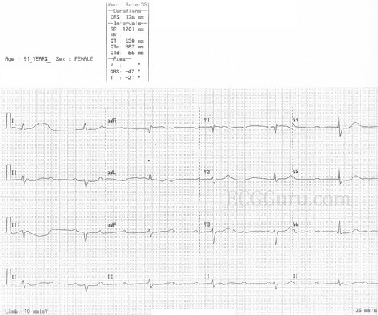
Instructors' Collection ECG: Complete Heart Block or High Grade AVB?

ECG Guru

If the patient is showing signs of poor perfusion, we would stop here and prepare to increase the rate with a temporary pacemaker (transvenous or transcutaneous). The ventricular rate is around 35 bpm, and regular. Why is the rate so slow? There is no P wave in front of each QRS, so this is not sinus bradycardia.

EKG/ECG 98
Insiders

Sign Up for our Newsletter

This site is protected by reCAPTCHA and the Google Privacy Policy and Terms of Service apply.

Trending Sources

article thumbnail

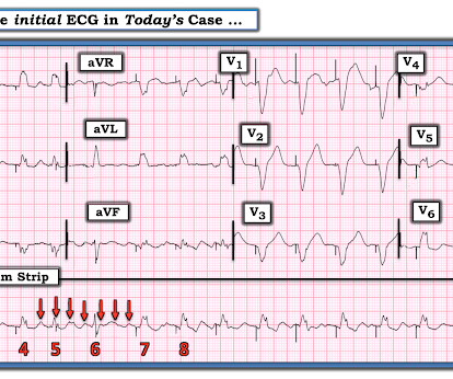
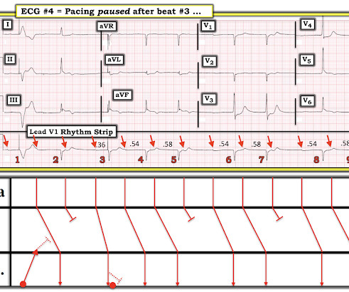
Emergency (ED placement) Transvenous Pacer appears to be working perfectly. What might go wrong?

Dr. Smith's ECG Blog

A transvenous pacer was placed in the Emergency Department by the emergency physicians. Asynchronous pacing seems like the safe setting, EXCEPT that it means that the pacemaker can trigger on the T-wave, which can result in Ventricular Fibrillation. PEARL #1: Most of the time the pacemaker is right!

EKG/ECG 52
article thumbnail

Grand Rounds Recap 1.10.24

Taming the SRU

Often AE-ILD is idiopathic, but treatable causes must be excluded (PNA, PE, volume overload) Treatment for AE-ILD should include antibiotics for CAP coverage (specifically including azithromycin), steroids, and respiratory support; consider opportunistic infection if immunosuppressed as well as diuresis as needed for euvolemia HFNC should be favored (..)

article thumbnail

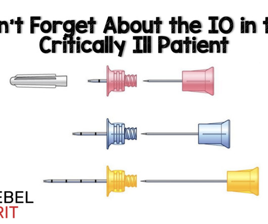
Don’t Forget About the IO in the Critically Ill Patient

RebelEM

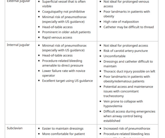
The “crash” or “dirty” central line, which forgoes strict sterile precautions, should be the last consideration in resuscitation as it increases risks for bloodstream infections, has more associated complications, and takes a potential access site required for ECMO cannulation, transvenous pacemaker, or other cardiac devices.

article thumbnail

Grand Rounds Recap 2.7.24

Taming the SRU

Location of the block will determine the morphology of the QRS (as a higher block may have a narrow QRS with a rate of 40-60bpm) Evaluation in the ED: basic labs including BMP and troponin, EKG, bedside echo, CXR Management: Atropine: push-dose 0.5-1mg,

article thumbnail

A woman in her 70s with bradycardia and hypotension

Dr. Smith's ECG Blog

She was intubated and a transvenous pacemaker was inserted with good capture. Figure-3: ECG #3 — recorded after ~30 minutes in the ED, after placement of a transvenous pacemaker ( See text ). Atropine produced no response. The patient was placed on pressors and transcutaneous pads (which failed to capture).

EKG/ECG 52