article thumbnail

Case 20 – Stethoscopically occult pneumonopathies

Urgent Care Ultrasounds

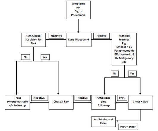
On examination she appeared well, t = 38.0. She had had a cough for the past month but has been otherwise well. She appeared well. Left PLAPS point Despite the obvious consolidation on ultrasound the CXR is clear. In all three there was clear ultrasound evidence of pneumonia.

article thumbnail

Red Leg in the Heartland of America: A Rural Physician’s Approach to the Patient with a Potential DVT

EMDocs

The facility does not have ultrasound (US) availability at the time of the patient’s presentation, as the sonographer comes to the hospital only 2 days a week. First, examine the patient and risk-stratify them for the diagnosis of DVT before deciding on the clinical utility of an ultrasound. What findings make a DVT more likely?

Insiders

Sign Up for our Newsletter

This site is protected by reCAPTCHA and the Google Privacy Policy and Terms of Service apply.

Trending Sources

article thumbnail

Case 19 – Through the looking glass

Urgent Care Ultrasounds

A window to your heart… A woman in her 40’s presented to the urgent care clinic with a four day history of cough and fevers following an upper respiratory tract infection the week prior. On examination she appeared well. She had noticed some mild SOB on exertion over the past few days when climbing stairs.

article thumbnail

Three normal high sensitivity troponins over 4 hours with a "normal ECG"

Dr. Smith's ECG Blog

Written by Willy Frick A 46 year old man with a history of type 2 diabetes mellitus presented to urgent care with complaint of "chest burning." In fact, all the T waves are more upright than his prior urgent care ECG. The distal LCx is seen, and the OM is not well visualized here. The following ECG was obtained.

EKG/ECG 92
article thumbnail

Case 15 – Lingulitis

Urgent Care Ultrasounds

On examination he appeared well, t = 38.0, A lung ultrasound was performed just to be sure… left base posteriorly Following the B lines around to the left precordium Disposition and Discussion An area of consolidation with air bronchograms is present in the left upper lobe. So a simple case of the flu?

article thumbnail

Case 16 – Come out, come out, wherever you are

Urgent Care Ultrasounds

The importance of patient positioning, probe position and patience, for the detection of small pneumothoraces Ultrasound to rule out a pneumothorax is easy! Well, maybe. If you are using ultrasound in the primary care setting as a rule out test, you really don’t want to miss any pneumothorax, however small.