Remove ecg benign-early-repolarization
article thumbnail

ECG Blog #365 — A 30yo with Pericarditis.

Ken Grauer, MD

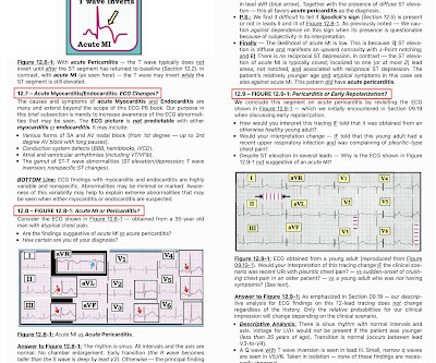
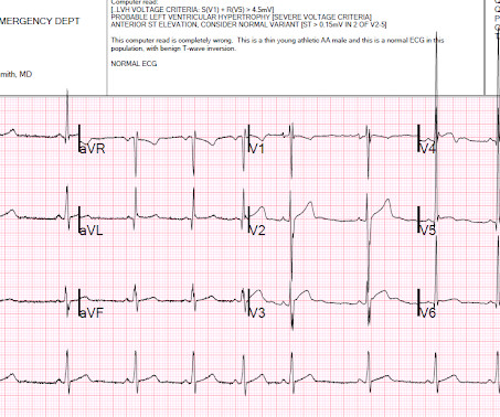
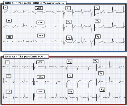
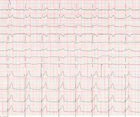
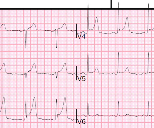
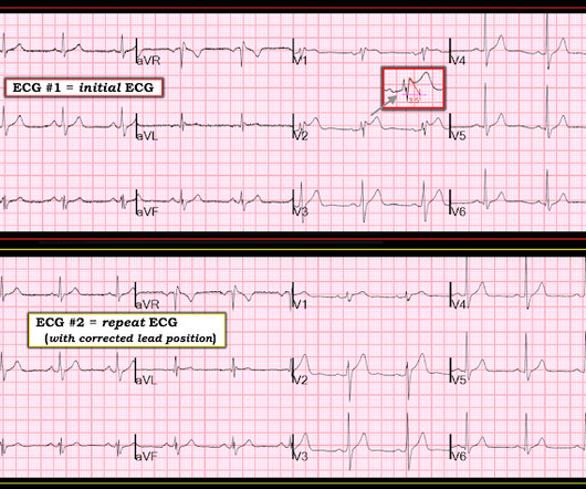
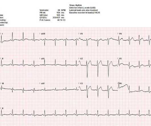
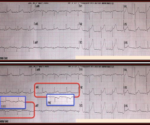
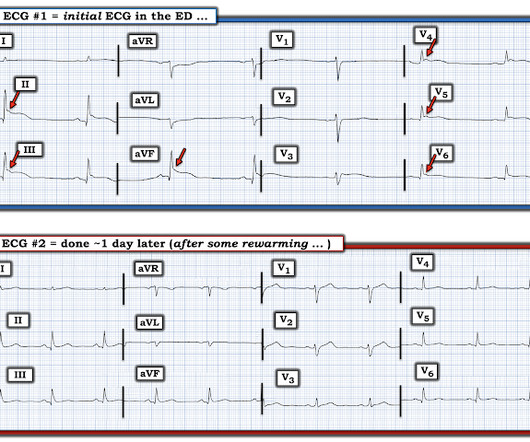
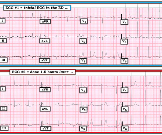
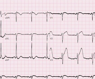
The ECG in Figure-1 was obtained from a previously healthy 30-ish year old man — who presented to medical attention for vasovagal syncope. Based on this initial ECG — the patient was transferred to a PCI-capable center: Do YOU agree with the need for transfer? Figure-1: The initial ECG in today's case.

EKG/ECG 195
article thumbnail

ECG Blog #409 — Every-Other-Beat.

Ken Grauer, MD

The ECG in Figure-1 — was obtained from a patient with palpitations. Figure-1: The initial ECG in today's case. My Interpretation of the ECG in Figure-1: Since the patient is hemodynamically stable — there is time for systematic assessment of the rhythm. Figure-2: I've labeled the initial ECG in today's case. Figure-2 ).

EKG/ECG 430
Insiders

Sign Up for our Newsletter

This site is protected by reCAPTCHA and the Google Privacy Policy and Terms of Service apply.

Trending Sources

article thumbnail

Acute OMI or "Benign" Early Repolarization?

Dr. Smith's ECG Blog

The following ECG was obtained. Note that the machine read is "normal sinus rhythm, normal ECG." ECG 1 What do you think? I sent this ECG to Dr. Smith and Dr. Meyers with no clinical context. Smith comment: this troponin alone should be enough data to activate the cath lab, regardless of the ECG. <0.049 ng/mL).

EKG/ECG 101
article thumbnail

Early repol or anterior OMI?

Dr. Smith's ECG Blog

Chest Pain – Benign Early Repol or OMI? Written by Destiny Folk, MD, Adam Engberg, MD, and Vitaliy Belyshev MD A man in his early 60s with a past medical history of hypertension, type 2 diabetes, obesity, and hyperlipidemia presented to the emergency department for evaluation of chest pain.

EKG/ECG 93
article thumbnail

Chest pain with 2 serial ECGs, with dynamic change, texted to me

Dr. Smith's ECG Blog

Queen: #1: NOT OMI, HIGH CONFIDENCE Queen: #2: NOT OMI, HIGH CONFIDENCE ECG 1 Interpretation: there is terminal T-wave in V3-V6. this is classic Benign T-wave Inversion. See 2 dozen examples here: Understanding this pathognomonic ECG would have greatly benefitted the patient. We just give it ECGs and tell it what they represent.

EKG/ECG 52
article thumbnail

Two patients with chest pain, with QRS obscured: which was STEMI positive, and which had Occlusion MI?

Dr. Smith's ECG Blog

Patient 1 (ECG on the left) was a 45 year-old male, and patient 2 (ECG is on the right) was a 70 year-old male. Using T wave amplitude, can you tell which ECG has hyperacute T waves? What other parts of the ECG would you like to see, and how will you use this to refine your interpretation?

EKG/ECG 86
article thumbnail

Concerning EKG with a Non-obstructive angiogram. What happened?

Dr. Smith's ECG Blog

That's because this patient has clinical, ECG and Troponin evidence that an acute event did in fact occur ( ie, a more than doubling of Troponin in the context of a changing pattern of CP that correlates temporally with "dynamic" ECG changes — therefore strongly suggesting transient acute occlusion regardless of cath findings in today's case ).

EKG/ECG 104