Remove ecg junctional-tachycardia
article thumbnail

Ventricular Fibrillation, ICD, LBBB, QRS of 210 ms, Positive Smith Modified Sgarbossa Criteria, and Pacemaker-Mediated Tachycardia

Dr. Smith's ECG Blog

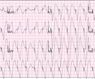
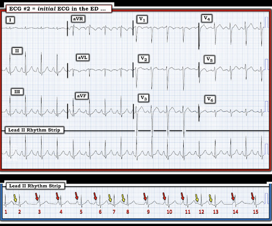
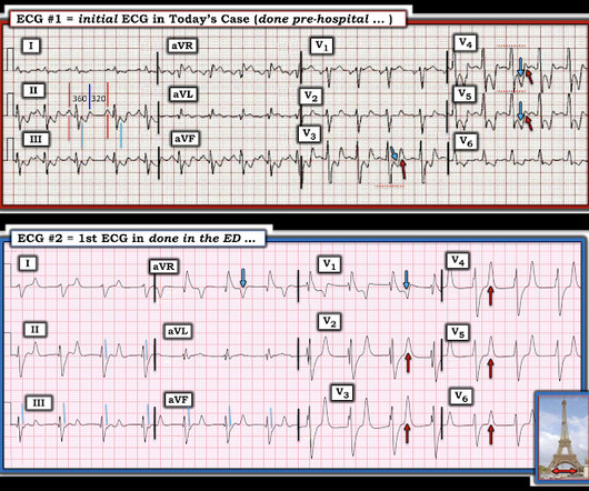
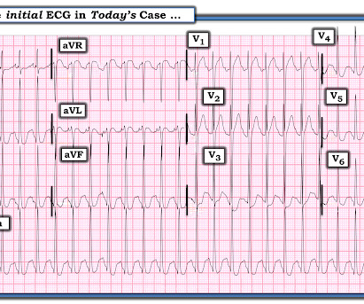
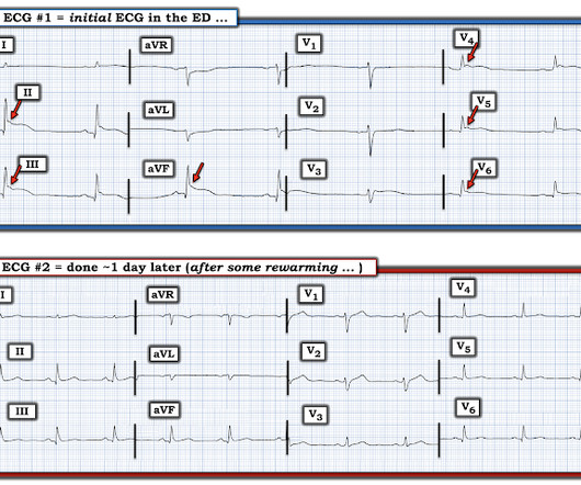
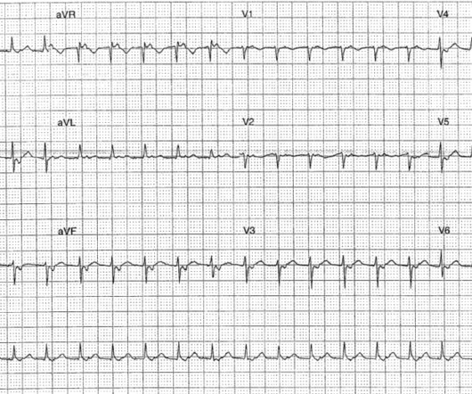
Here is the initial ED ECG. Then I always look to see if the initial deflection of the QRS has a lot of voltage change per change in time (seen in tachycardias that are initiated from above the ventricle because the propagate through fast conducting purkinje fiber. Tachycardia exaggerates ST Elevation in LBBB and Paced rhythm 5.

EKG/ECG 114
article thumbnail

A man in his 40s with acute chest pain. What do you think?

Dr. Smith's ECG Blog

Triage ECG: What do you think? It's a very "fun" ECG, with initial ectopic atrial tachycardia (negative P waves in inferior leads conducting 1:1 with the QRSs), followed by spontaneous resolution to sinus rhythm. Sudden CP and SOB with Inferior ST Elevation and in STE in V1. Is it inferior and RV OMI?

EKG/ECG 103
Insiders

Sign Up for our Newsletter

This site is protected by reCAPTCHA and the Google Privacy Policy and Terms of Service apply.

Trending Sources

article thumbnail

An Intriguing Rhythm: Who Belongs to Whom?

Dr. Smith's ECG Blog

Figure-1: The initial ECG in today's case. ( To improve visualization — I've digitized the original ECG using PMcardio ). Since the QRS complex for each of these non-sinus-conducted beats is narrow — beats #3,4; 9; 14 and 15 must be junctional beats. NOTE: It is unclear if beat #5 is junctional or sinus-conducted.

EKG/ECG 71
article thumbnail

ECG Blog #391 — Asymptomatic but Irregular.

Ken Grauer, MD

He was not symptomatic with the ECG shown in Figure-1. How would YOU interpret this ECG? Extra Credit ( which is a HINT to the Answer! ): How many beats are recorded on the ECG in Figure-1 ? Figure-1: The initial ECG in today’s case. ( To improve visualization — I've digitized the original ECG using PMcardio ).

EKG/ECG 195
article thumbnail

What is the rhythm?

Dr. Smith's ECG Blog

Here is the only prehospital 12-lead: Sinus tachycardia, somewhat wide QRS, Ischemia Here is the first ED ECG: What is the rhythm here? This ECG is pathognomonic of hyperkalemia , with wide QRS, very SHARPLY peaked T-waves, flat ST segments, RBBB pattern and large R-wave in aVR. Is this an accelerated Junctional rhythm?

EKG/ECG 76
article thumbnail

A young man with tachycardia. Should We Try Adenosine?

Dr. Smith's ECG Blog

An ECG was recorded: What do you think? There is a regular narrow complex tachycardia. Thus, it is supraventricular tachycardia. It is important to remember that SVT includes Sinus Tachycardia! Sometimes even Wide Complex Tachycardia is Sinus. A very fast narrow complex tachycardia in an Infant 3.

EKG/ECG 78
article thumbnail

What do you suspect from this ECG in this 40-something with SOB and Chest pain?

Dr. Smith's ECG Blog

I was reviewing ECGs for a study, and came across this one, and was able to get all the clinical information: What do you think? Medics recorded the above ECG and called a STEMI alert. The 2 ECGs that were recorded in today's case are insightful. The Queen diagnosed "OMI with high confidence" due to the ST Elevation in V1-V3.

EKG/ECG 68