Remove ecg left-posterior-fascicular-block
article thumbnail

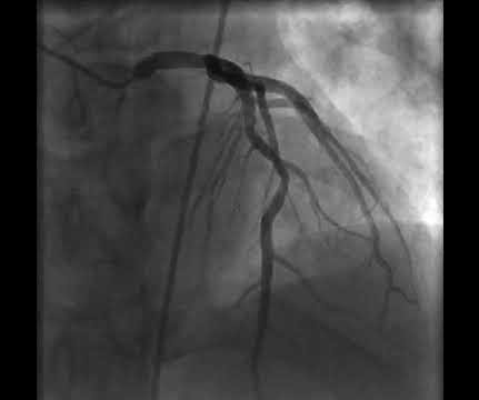
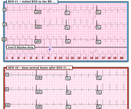
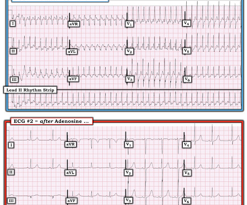
A 50-something with chest pain. Is there OMI? And what is the rhythm?

Dr. Smith's ECG Blog

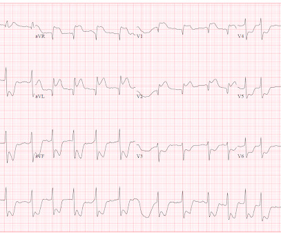
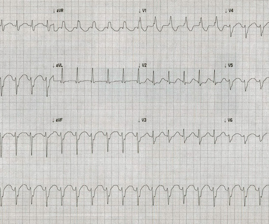
His ECG is shown: What do you think? The history thus far is highly suggestive of OMI, so we must study the ECG very closely to see if we can confirm this. The Queen of Hearts does not care about rhythm analysis, she simply looks at the ECG and decides whether it represents OMI or not. Due to the precordial swirl Dr.

EKG/ECG 99
article thumbnail

Sudden shock with a Nasty looking ECG. What is it?

Dr. Smith's ECG Blog

There were 2 prehospital ECGs: What do you think? There is "shark fin" in I and aVL, which is due to a combination of a large R-wave due to left anterior fascicular block plus downsloping ST elevation due to OMI. Moreover, left main occlusion often presents near death. Posterior and high lateral OMI.

Shock 98
Insiders

Sign Up for our Newsletter

This site is protected by reCAPTCHA and the Google Privacy Policy and Terms of Service apply.

Trending Sources

article thumbnail

Wide complex tachycardia and hypotension in a 50-something with h/o cardiomyopathy -- what is it?

Dr. Smith's ECG Blog

Here is the ED ECG: What do you think? Thus, it is probably SVT with aberrancy (RBBB + LAFB) or it is posterior fascicular VT (which starts in the posterior fascicle and therefore also has a fast initial depolarization nearly identical to RBBB + LAFB). Analysis: there is a wide complex tachycardia. It is regular.

EKG/ECG 52
article thumbnail

Massive Transfusion for Motorcycle Collision with Hemorrhage, Troponin Elevated.

Dr. Smith's ECG Blog

This ECG was done in a middle aged woman who was in a motor vehicle collision in which her vehicle "T-boned" another, so there was trauma to the anterior chest. ng/mL, and an ECG was recorded: There are no P-waves visible. --Sinoventricular rhythm RBBB and LAFB morphology. The estimated left ventricular ejection fraction is 76 %.

article thumbnail

Young Man with a Heart Rate of 257. What is it and how to manage?

Dr. Smith's ECG Blog

The Differential Diagnosis is: SVT with aberrancy(#) [AVNRT vs. WPW (also called AVRT*)] Atrial flutter with 1:1 conduction, with aberrancy VT coming from the anterior fascicle ( fascicular VT )@ *AVRT = AV Reciprocating Tachycardia (Tachycardic loop that uses both the AV node and an accessory pathway. This confirms SVT, either AVNRT or WPW.

EKG/ECG 52
article thumbnail

How does Acute Total Left Main Coronary occlusion present on the ECG?

Dr. Smith's ECG Blog

Post by Smith and Meyers Sam Ghali ( [link] ) just asked me (Smith): "Steve, do left main coronary artery *occlusions* (actual ones with transmural ischemia) have ST Depression or ST Elevation in aVR?" The pattern seen may, or may not, be unique to left main occlusion. All are, however, clearly massive STEMI.

EKG/ECG 52
article thumbnail

Wide Complex Tachycardia. What is the Diagnosis?

Dr. Smith's ECG Blog

On arrival, here is his initial ED ECG, and is identical to the prehospital ECG and at the same rate: There is a regular, wide complex. The wide complex has a right bundle branch and left anterior fascicular block morphology. Alternatively, it could be posterior fascicular ventricular tachycardia.

EKG/ECG 52