Remove ecg non-conducted-pacs
article thumbnail

ECG Blog #429 — Mobitz I or Mobitz II?

Ken Grauer, MD

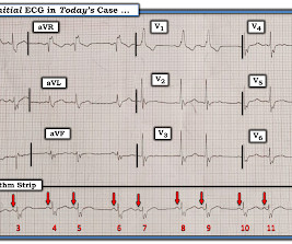
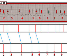
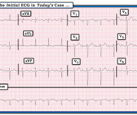
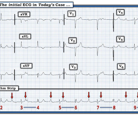
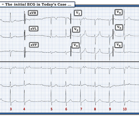
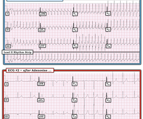
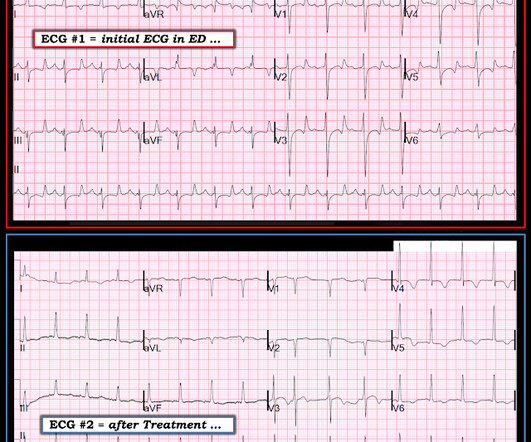
The 12-lead ECG and long lead II rhythms shown in Figure-1 — was obtained from an older man with a recent history of “easy fatiguability” and a presyncopal episode. QUESTIONS: How would YOU interpret the ECG in Figure-1 ? Figure-1: The initial ECG in today’s case. I outline my approach for doing so below.

EKG/ECG 263
article thumbnail

ECG Blog #385 — This Patient Arrested Soon After

Ken Grauer, MD

The ECG in Figure-1 was obtained from a 60-year old woman — who presented to the ED ( E mergency D epartment ) for “palpitations”. Her vital signs were normal at the time this triage ECG was recorded. How would YOU interpret the ECG in Figure-1 ? Do you see any indication on this ECG of WHY this patient was about to arrest?

EKG/ECG 195
Insiders

Sign Up for our Newsletter

This site is protected by reCAPTCHA and the Google Privacy Policy and Terms of Service apply.

Trending Sources

article thumbnail

ECG Blog #391 — Asymptomatic but Irregular.

Ken Grauer, MD

He was not symptomatic with the ECG shown in Figure-1. How would YOU interpret this ECG? Extra Credit ( which is a HINT to the Answer! ): How many beats are recorded on the ECG in Figure-1 ? Figure-1: The initial ECG in today’s case. ( To improve visualization — I've digitized the original ECG using PMcardio ).

EKG/ECG 195
article thumbnail

ECG Blog #389 — A Quote from Sherlock Holmes

Ken Grauer, MD

What is unusual about this conduction disturbance? Are Any P Waves in Figure-2 Conducting? The Question that now arises is — Are any of the P waves that we see in Figure-2 being conducted to the ventricles? Her vital signs were stable — and she did not have an acute abdomen. QUESTIONS: Is there complete AV block?

EKG/ECG 370
article thumbnail

ECG Blog #398 — Uncontrolled Graves Disease.

Ken Grauer, MD

The ECG in Figure-1 was obtained from a middle-aged woman who presented with "palpitations". NOTE: Although the most common cause of unexpected pauses in a rhythm is the occurrence of one or more blocked PACs — I do not see distortions in any ST-T wave of this single-lead rhythm strip that might be consistent with blocked PACs.

EKG/ECG 384
article thumbnail

ECG Blog #416 — Is the Rhythm and ECG related?

Ken Grauer, MD

Imagine the only information provided for the ECG in Figure-1 — is that it was obtained from a 60-year old man with new CP ( C hest P ain ). QUESTIONS: In view of this brief history — How would YOU interpret this ECG in Figure-1 ? Is the cardiac rhythm related to the 12-lead ECG? Figure-1: The initial ECG in today’s case.

EKG/ECG 255
article thumbnail

ECG Blog #397 — An Unusually Long Cycle?

Ken Grauer, MD

You are asked to interpret the ECG in Figure-1. What is the rhythm in ECG #1 ? MY Thoughts on the ECG in Figure-1: I routinely begin assessment of each 12-lead ECG I encounter — with interpretation of the rhythm. To do this — I apply the P s, Q s, 3 R Approach ( See ECG Blog #185 — for review of my system ).

EKG/ECG 239