Remove lv
article thumbnail

Tasty Morsels of Critical Care 074 | Dynamic LV outflow tract obstruction

Emergency Medicine Ireland

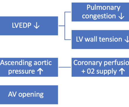
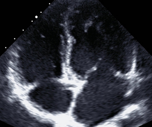
Today we’re going to verge into challenging territory for an audio podcast in that we’re going to the discuss the very visual topic of dynamic LV outflow tract obstruction. The LV receives less than usual volume to stretch it and the low afterload makes it incredibly easy for the LV to empty itself of this load.

Sepsis 52
article thumbnail

Quantitative LV Function in PEA

Ultrasound Gel

Current practice for many is to assess for the binary presence or absence of cardiac activity, but these audacious authors suggest that a higher LV systolic function could be associated with a greater likelihood of return of spontaneous circulation! [link] [link]

Insiders

Sign Up for our Newsletter

This site is protected by reCAPTCHA and the Google Privacy Policy and Terms of Service apply.

Trending Sources

article thumbnail

POCUS Cases 8 – LV Dysfunction

Emergency Medicine Cases

In this POCUS Cases video Dr. Rob Simard reviews the literature on accuracy of identifying LV dysfunction on POCUS by non-radiologists, the steps in assessing LV dysfunction, and cautions us when it comes to patients with chronic LV dysfunction.

article thumbnail

Mallia – Critical Care Ultrasonographic Assessment of the LV and RV

University of Maryland CC Project

Mallia, Assistant Professor of Emergency Medicine and Associate Program Director of the Pulmonary and Emergency Medicine-Critical Care Fellowship at MedStar Washington Hospital presents on critical care ultrasound for the assessment of the LV and RV as part of the DC5 lecture series. Dr. Anantha K.

article thumbnail

POCUS findings of hemodynamically unstable PE with cardiac arrest

EMDocs

At first pulse check, EPs ascertained the patient had no LV function (asystole), no pericardial effusion, but did recognize that the RV cavity appeared subtly larger than the LV cavity, indicating possible RV strain. SubX4 Asystole RV > LV. SubX2 Asystole RV > LV. SubX4 ROSC RV > LV. 10,11 Vid 1.

article thumbnail

Acute Dyspnea and Right Bundle Branch Block

Dr. Smith's ECG Blog

LV aneurysm has QS-waves, so this couldn't be LV aneurysm, right? RBBB makes it mandatory that there are R'-waves even in the presence of LV aneurysm. Additionally, it is very difficult to differentiate subacute reperfused OMI from LV aneurysm: both have Q-waves and inverted T-waves. This is HIGHLY suspicious for OMI.

EKG/ECG 101
article thumbnail

ECG Blog #427 — To Cath this Elderly Patient?

Ken Grauer, MD

PEARL # 3: It may sometimes be difficult to distinguish between active, ongoing infarction — vs LV aneurysm that developed following a prior infarction — vs superimposed new infarction, that occurs on top of prior infarction.

EKG/ECG 332