Remove ecg right-ventricular-mi
article thumbnail

ECG Blog #416 — Is the Rhythm and ECG related?

Ken Grauer, MD

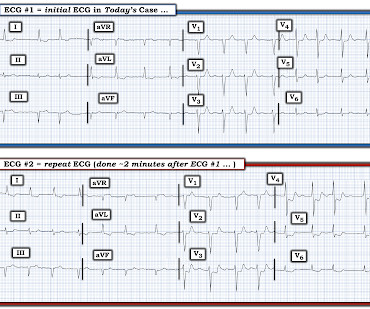
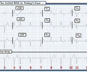
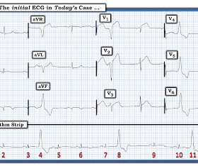
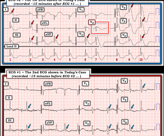
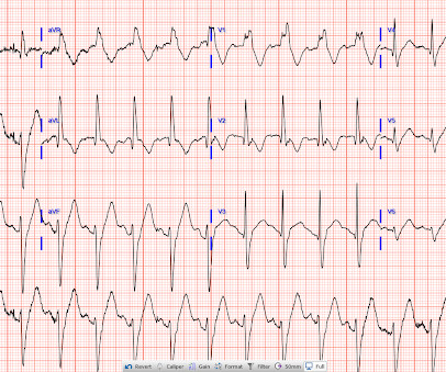
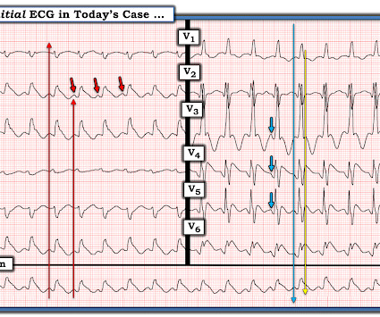
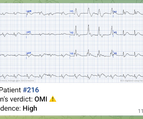
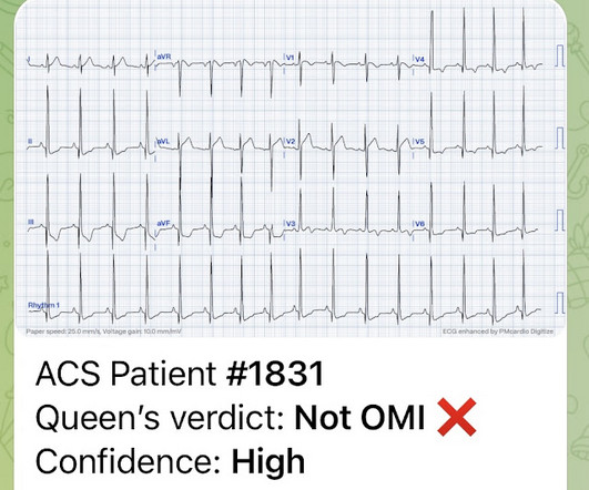
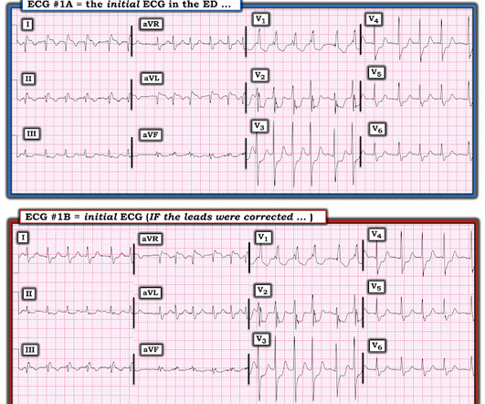
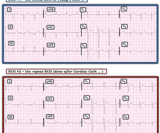
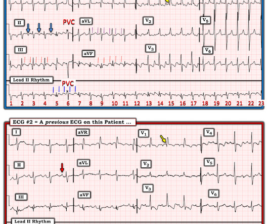
Imagine the only information provided for the ECG in Figure-1 — is that it was obtained from a 60-year old man with new CP ( C hest P ain ). QUESTIONS: In view of this brief history — How would YOU interpret this ECG in Figure-1 ? Is the cardiac rhythm related to the 12-lead ECG? Figure-1: The initial ECG in today’s case.

EKG/ECG 255
article thumbnail

ECG Blog #387 — 2 Minutes Later.

Ken Grauer, MD

The ECG in Figure-1 was obtained from an elderly man with a history of coronary disease — who contacted EMS for "burning" chest discomfort that woke him at 3am. Some amount of time passed at home — during which his chest discomfort persisted The patient was hemodynamically stable at the time he was seen by EMS, when ECG #1 was recorded.

EKG/ECG 195
Insiders

Sign Up for our Newsletter

This site is protected by reCAPTCHA and the Google Privacy Policy and Terms of Service apply.

Trending Sources

article thumbnail

SGEM#428: Don’t Worry, Be Happy – The Safety of Nitroglycerin Administration in RVMI

The Skeptics' Guide to EM

Adverse events from nitrate administration during right ventricular myocardial infarction: a systematic review and meta-analysis. Adverse events from nitrate administration during right ventricular myocardial infarction: a systematic review and meta-analysis. In all comers with MI, the rate is probably closer to <3%.

article thumbnail

ECG Blog #373 — 86yo and this Rhythm.

Ken Grauer, MD

The 12-lead ECG and long lead II rhythm strip in Figure-1 was obtained from an 86-year old man — who presented to the ED ( E mergency D epartment ) with presyncope. How would YOU interpret the ECG in Figure-1 ? Figure-1: The initial ECG in today's case — obtained from an 86-year old man with presyncope, but no chest pain. (

EKG/ECG 195
article thumbnail

Chest pain and shock: Is there a right ventricular OMI on this ECG? And should he undergo trancutaneous pacing?

Dr. Smith's ECG Blog

His prehospital ECG was diagnostic of inferior posterior OMI. Here is his ED ECG: There is bradycardia with a junctional escape. Is there also a right ventricular OMI (RV OMI, or RV MI)? STD in V2 "pulls down" the ST segment in V1 thus negating any sign of RV MI. He appeared gray in color, with cool skin.

Shock 71
article thumbnail

Chest pain with anterior ST depression: look what happens if you use posterior leads.

Dr. Smith's ECG Blog

There’s atrial flutter with controlled ventricular response, a non-specific intra-ventricular conduction delay, borderline right axis, normal R wave progression and normal voltages. Here's the prior ECG: This confirms thew anterior ST depression is new. What do you think? Do you need posterior leads?

EKG/ECG 100
article thumbnail

ECG Blog #359 — How Many Beats in the 12-Lead?

Ken Grauer, MD

The ECG in Figure-1 was obtained from a previously healthy older man — who complained of chest pain and “lightheadedness” while this tracing was recorded. In view of this history — How would YOU interpret the ECG in Figure-1 ? To improve visualization — I've digitized the original ECG using PMcardio ). He was not hypotensive.

EKG/ECG 195