article thumbnail

Chest pain, resolved. Does it need emergent cath lab activation (some controversy here)? And much much more.

Dr. Smith's ECG Blog

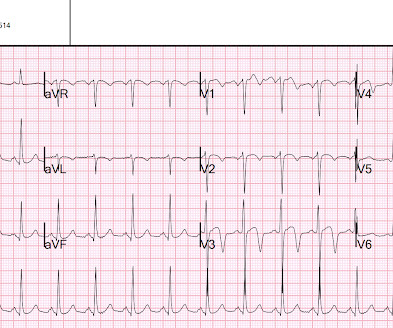
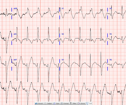
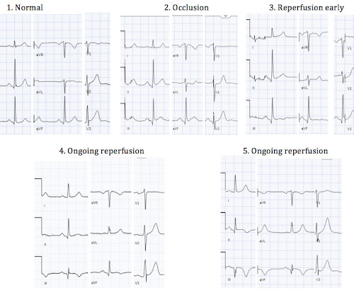
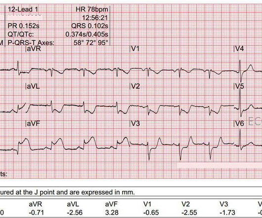
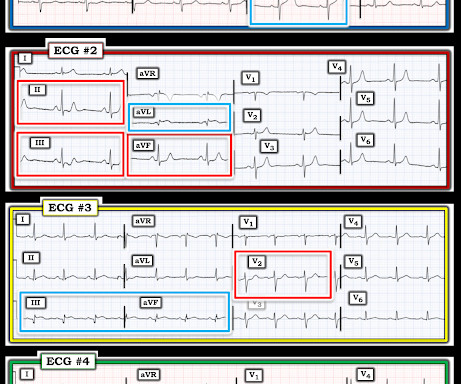
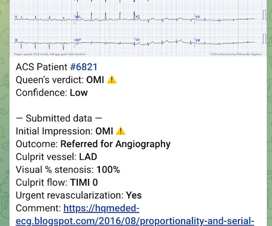
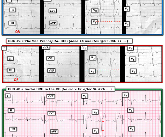
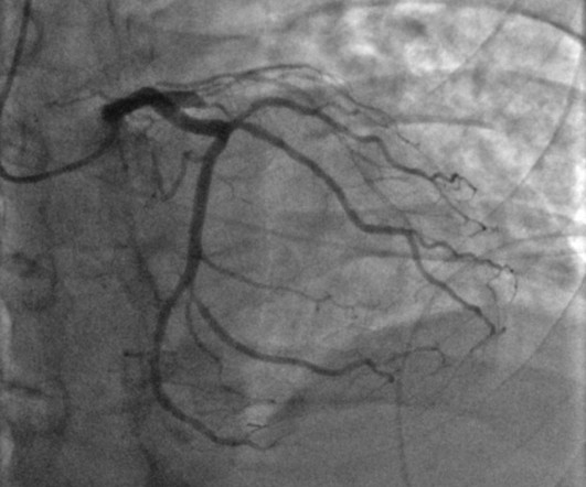
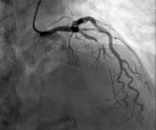
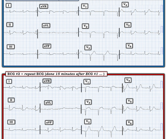
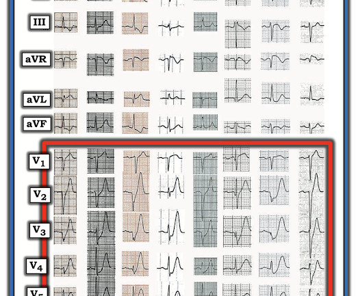
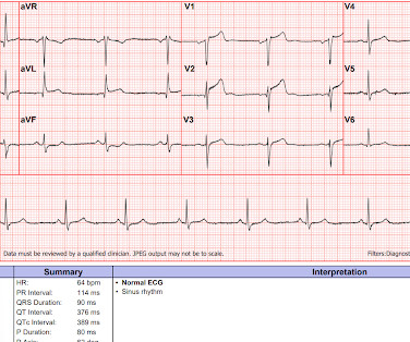
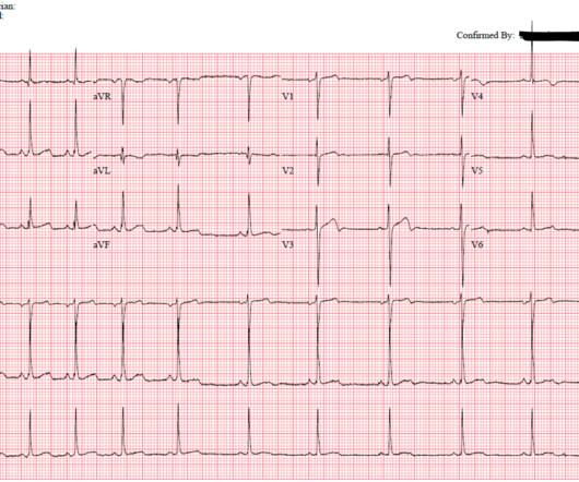
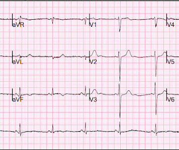
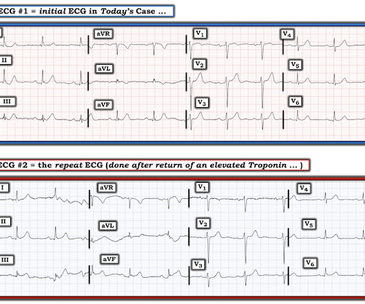
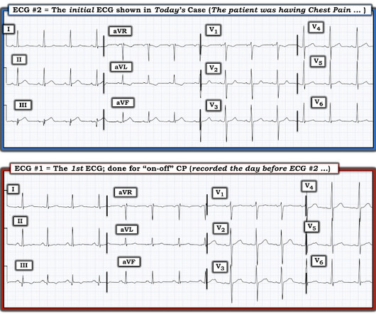
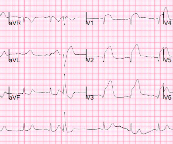
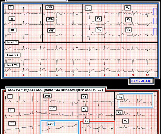
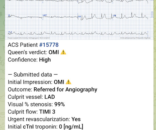
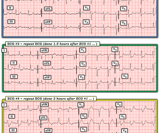
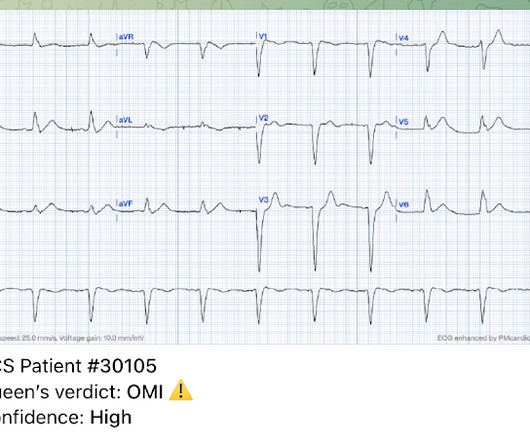
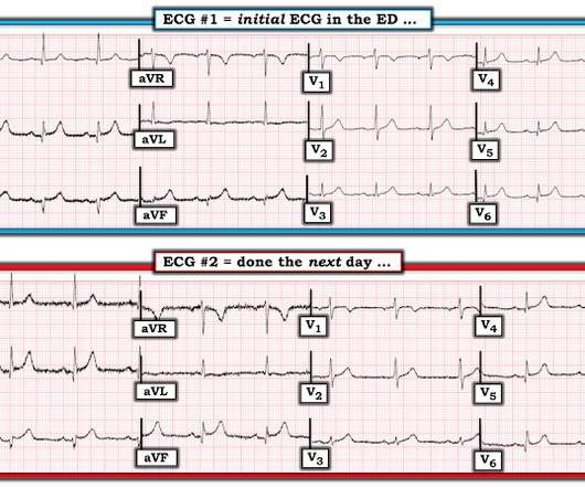
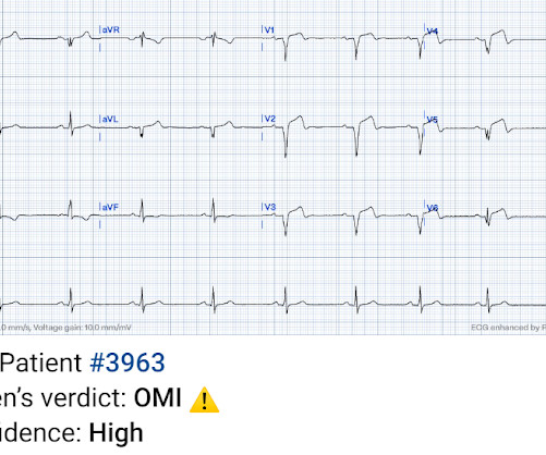
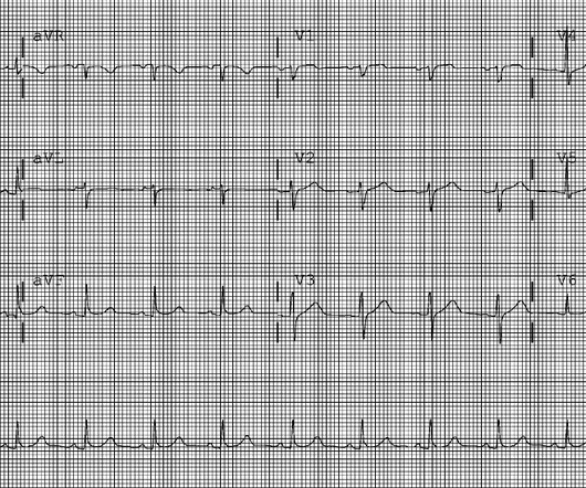
EKG from triage: Here is his previous ECG: Normal ST Elevation Resident's interpretation: Reperfusion pattern/Wellens' with biphasic T waves in V2 and V3, and in comparison to an EKG in 2020 this is new. Repeat EKG: Resident interpretation: ST elevation in V2 significantly different than his previous EKG.

EKG/ECG 110
article thumbnail

A teenager involved in a motor vehicle collision with abnormal ECG

Dr. Smith's ECG Blog

Here is his initial ECG around 1330: What do you think? The ECG shows sinus tachycardia with RBBB and LAFB, without clear additional superimposed signs of ischemia. Initial high sensitivity troponin I: 3,830 ng/L (URL 20 ng/L for men) 1445: Similar to initial ECG. He was intubated for altered mental status.

EKG/ECG 96
Insiders

Sign Up for our Newsletter

This site is protected by reCAPTCHA and the Google Privacy Policy and Terms of Service apply.

Trending Sources

article thumbnail

ECG Blog #366 — Diltiazem didn't work.

Ken Grauer, MD

The ECG and long lead II rhythm strip in Figure-1 — was obtained from a COVID positive patient with persistent tachycardia not responding to Diltiazem. Figure-1: The initial ECG — obtained from a patient with persistent tachycardia. ( To improve visualization — I've digitized the original ECG using PMcardio ).

EKG/ECG 195
article thumbnail

Occlusion myocardial infarction is a clinical diagnosis

Dr. Smith's ECG Blog

Willy is a cardiology fellow with a keen interest in the ECG in OMI. If an immediate EKG was obtained, it was not saved in the medical record. The first available EKG was recorded just after midnight, presumably around the time the result of the troponin came to clinical attention. The patient said, "I just don't feel good."

EKG/ECG 113
article thumbnail

Chest pain and a computer ‘normal’ ECG. Therefore, there is no need for a physician to look at this ECG.

Dr. Smith's ECG Blog

Below is the triage ECG, with a computer interpretation (Marquette 12 SL) of “normal” which was confirmed by the over-reading cardiologist. Should this patient continue to stay in the waiting room, without interruption of the physician to interpret the ECG, because the computer interpretation is normal? What do you think?

EKG/ECG 91
article thumbnail

Is there OMI on this ECG?

Dr. Smith's ECG Blog

The patient with no prior cardiac history presented in the middle of the night with acute chest pain, and had this ECG recorded during active pain: I did not see any ischemia on this electrocardiogram. I think the ECG is normal. OMI is a clinical diagnosis that includes the ECG. This is a case I had quite a while back.

EKG/ECG 92
article thumbnail

Instructors' Collection ECG: Inferior Posterior M.I.

ECG Guru

This is a "classic" ECG of very good quality for you to use in a classroom setting. The ECG: This ECG could be considered "classic" for an inferior wall ST elevation M.I. The ECG: This ECG could be considered "classic" for an inferior wall ST elevation M.I. He is pale, cool, and diaphoretic.

EKG/ECG 98