article thumbnail

Concerning EKG with a Non-obstructive angiogram. What happened?

Dr. Smith's ECG Blog

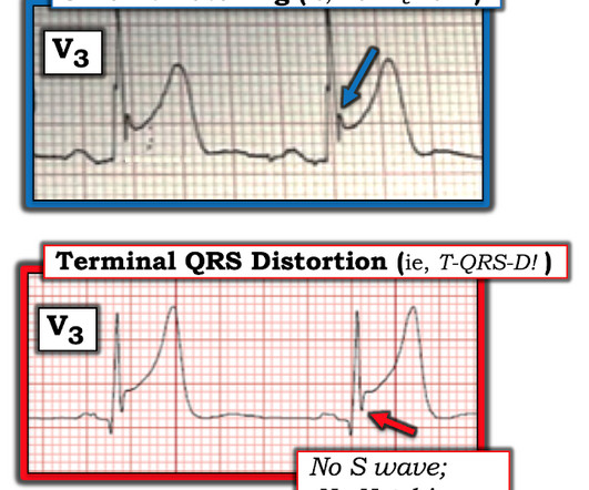
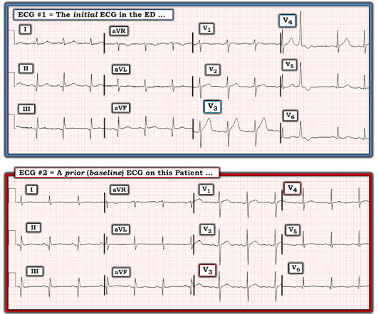
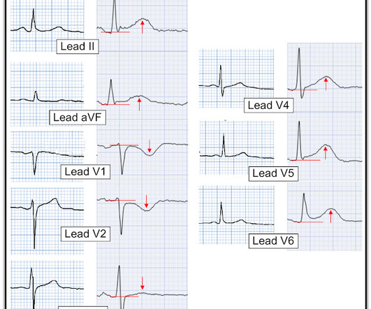
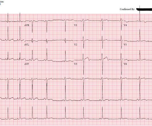
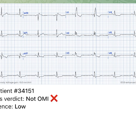
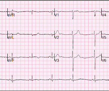
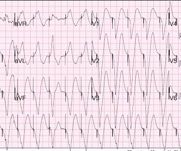
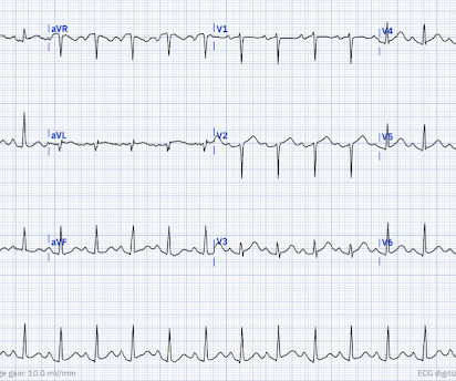
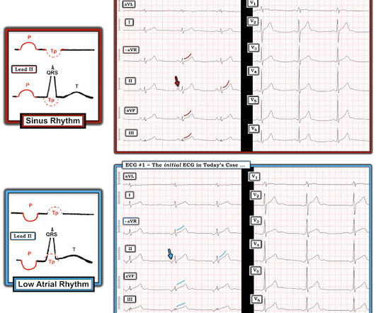
It appears EMS obtained two EKGs, but unfortunately these were not saved in the medical record. The EMS crew was only BLS certified, so EKG interpretation is not within their scope of practice. The patient arrived just after 10 AM, and the following EKG was obtained. There are no further EKGs or troponin measurements.

EKG/ECG 107
article thumbnail

Chest pain, resolved. Does it need emergent cath lab activation (some controversy here)? And much much more.

Dr. Smith's ECG Blog

EKG from triage: Here is his previous ECG: Normal ST Elevation Resident's interpretation: Reperfusion pattern/Wellens' with biphasic T waves in V2 and V3, and in comparison to an EKG in 2020 this is new. Bedside ultrasound with no apparent wall motion abnormalities, no pericardial effusion, no right heart strain.

EKG/ECG 110
Insiders

Sign Up for our Newsletter

This site is protected by reCAPTCHA and the Google Privacy Policy and Terms of Service apply.

Trending Sources

article thumbnail

Expert human ECG interpretation and/or the Queen of Hearts could have saved this patient's anterior wall

Dr. Smith's ECG Blog

He had active chest pain at the time of triage at 0137 at night, with this triage ECG: What do you think? I sent this ECG, without any text at all, to Dr. Smith, and he replied: "LAD OMI with low certainty. Back to the case: Unfortunately, the ECG was not understood by the provider. V3 is the one that is convincing."

EKG/ECG 117
article thumbnail

The SPEED Protocol: Using Ultrasound To Detect Acute Aortic Dissection

ACEP Now

His vital signs are normal and the ECG does not demonstrate a myocardial infarction. While waiting for laboratory investigations, including troponin and d-dimer, you wonder if a quick point-of-care ultrasound (POCUS) examination looking for three sonographic findings could help determine the likelihood of this being an AoD.

article thumbnail

Four patients with chest pain and ‘normal’ ECG: can you trust the computer interpretation?

Dr. Smith's ECG Blog

All initial ECGs were labeled ‘normal’ or ‘otherwise normal’ by the computer interpretation, and below are the ECGs with the final cardiology interpretation. 1-3] But these studies were very short duration and used cardiology interpretation of ECGs or emergent angiography rather than patient outcomes.

EKG/ECG 111
article thumbnail

Three normal high sensitivity troponins over 4 hours with a "normal ECG"

Dr. Smith's ECG Blog

The following ECG was obtained. ECG 1 What do you think? The ECG shows sinus bradycardia but is otherwise normal. There is TWI in lead III, but this can be seen in normal ECGs. The following ECG was obtained around midnight. The machine read was "Normal sinus rhythm, normal ECG." ECG 2 What do you think?

EKG/ECG 95
article thumbnail

Chest pain in a 30-something: Is it Normal variant STE or OMI? Get the prior ECG, and don't trust Point of Care troponin assays!

Dr. Smith's ECG Blog

Triage EKG: What do you think? A prior ECG was available for comparison. Prior EKG from 2 months ago was available: Let's put the precordial leads from the 2 ECGs side by side: Now you can really see the difference. Immediately after the second ECG was performed, the patient's pain resolved completely.

EKG/ECG 52