Remove ultrasound-of-the-month
article thumbnail

Ultrasound of the Month: Peritonsillar Abscess

Taming the SRU

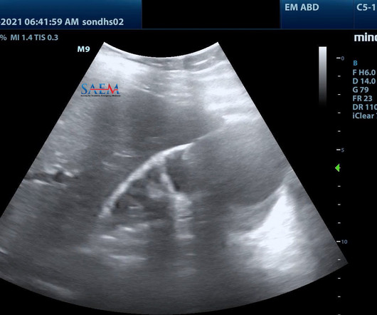
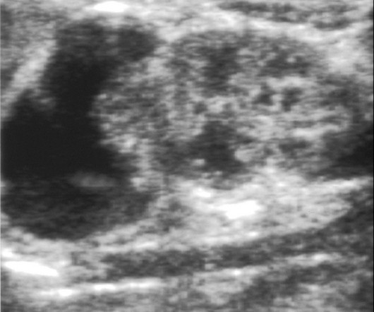
IMAGING WITH ULTRASOUND Peritonsillar abscess is one of the most common deep space infections of the head and neck contributing significantly to health care costs in the United States. THE CASE A female in her 20’s presented to the emergency department with concern for oral swelling. There was no trismus.

article thumbnail

Ultrasound of the Month: Gallbladder Perforation

Taming the SRU

A bedside right upper quadrant ultrasound was performed, and the images are below. THE CASE A male in his 60s presented to the emergency department for right upper quadrant abdominal pain. coli) empyema due to complicated pneumonia, and a prior perisplenic abscess requiring percutaneous drainage. The remainder of his exam was unremarkable.

Insiders

Sign Up for our Newsletter

This site is protected by reCAPTCHA and the Google Privacy Policy and Terms of Service apply.

Trending Sources

article thumbnail

Ultrasound of the Month: No Definitive Yolk Sac, No Definitive IUP!

Taming the SRU

She has taken a home pregnancy test which was positive, and her last menstrual period was over one month ago. She has taken a home pregnancy test which was positive, and her last menstrual period was over one month ago. A bedside ultrasound is completed to assess the location of the pregnancy.

article thumbnail

Ultrasound of the Month: Severe Hydronephrosis in a patient with Horseshoe Kidney

Taming the SRU

Given her age, history of nephrolithiasis, and frequent urinary tract infections, a bedside renal ultrasound was performed to assess for hydronephrosis due to concern her generalized weakness may be secondary to an infected stone or obstructive uropathy. She denies any fevers, abdominal pain, nausea, or vomiting. Images are seen below.

article thumbnail

Ultrasound of the Month: Can you use Pocus for pneumonia?

Downeast Emergency Medicine

Credits: Andrew fried, md A point-of-care lung ultrasound is performed, with the following findings on the left lung fields. Credits: Andrew fried, md A point-of-care lung ultrasound is performed, with the following findings on the left lung fields. Point of Care Ultrasound of the Left Lung Fields.

article thumbnail

Ultrasound of the Month: A Case of Unilateral Knee Swelling

Downeast Emergency Medicine

Bedside musculoskeletal ultrasound of his knees was performed. Credits: Andrew fried, md His left (normal) knee is shown below: Labeled combined still frames of the above ultrasound pointing out landmarks That should BE noted on every knee ultrasound. He has no numbness or tingling radiating down his leg.

article thumbnail

Ultrasound of the Month - Is your probe marker switched?

Downeast Emergency Medicine

Bedside chest ultrasound showed large bilateral pleural effusions. CASE PRESENTATION A 73 y.o. male with history of diastolic heart failure, type II DM, HTN, and peripheral vascular disease presented to the emergency department via EMS for evaluation of abnormal vital signs. He improved to the mid 90s with 4L nasal cannula.